Werfen Sie einen Blick auf alle unsere anstehenden Konferenzen, Kongresse, Messen, Webinare und Workshops und besuchen Sie uns bei einer unserer nächsten Veranstaltungen!
26
Feb
2025
Designing the Future: Novel Stem Cell Culture and RNA Tech
United Kingdom
•
Webinar
Filter articles
Tags
Produkte
Loading...
![Complete camera overview of EM grid recorded with 3 channels. Complete camera overview of EM grid recorded with 3 channels. Inserts displaying the positions, where superresolved 3D confocal images were recorded. 3D renderings of these positions are shown in the zoomed inserts. Fluorescence channels (nuclei by Hoechst, blue; mitochondria by MitoTracker Green, green; lipid Droplets by Bodipy and Crimson Beads, red). Width of a grid square is 90 ?m, width of a grid bar is 35 ?m. Samples kindly provided by Ievgeniia Zagoriy, Mahamid-Group, EMBL Heidelberg, Germany.](/fileadmin/_processed_/d/3/csm_Coral_Cryo_EM-Grid_Overview_scan_z-stacks_baf8cf6a6c.jpg)
From Bench to Beam: A Complete Correlative Cryo Light Microscopy Workflow
In the webinar entitled "A Multimodal Vitreous Crusade, a Cryo Correlative Workflow from Bench to Beam" a team of experts discusses the exciting world of correlative workflows for structural biology…
Loading...
![Transfection using the Uncommon Bio reprogramming system. Image acquired using the THUNDER Imager 3D Cell Culture with THUNDER Large Volume Computational Clearing (LVCC) applied. Image courtesy of Samuel East, Uncommon Bio. Transfection using the Uncommon Bio reprogramming system. Image acquired using the THUNDER Imager 3D Cell Culture with THUNDER Large Volume Computational Clearing (LVCC) applied. Image courtesy of Samuel East, Uncommon Bio.](/fileadmin/_processed_/a/8/csm_Transfection_using_Uncommon_Bio_reprogramming_system_07fe8407a3.jpg)
Designing the Future with Stem Cell and RNA Technology
Visionary biotech start-up Uncommon Bio is tackling one of the world’s biggest health challenges: food sustainability. In this webinar, Stem Cell Scientist Samuel East will show how they use RNA…
Loading...
![Developing embryos of different species at different stages during the elongation of their posterior body axis, from left to right in developmental time. Developing embryos of different species at different stages during the elongation of their posterior body axis, from left to right in developmental time. The labelled regions in red depict a region of undifferentiated cells called the tailbud, with the corresponding region generated from that tissue shaded in grey. Upper row: lamprey; middle row: catshark; bottom row, zebrafish. This figure has been adapted from the following publication: Steventon, B., Duarte, F., Lagadec, R., Mazan, S., Nicolas, J.-F., & Hirsinger, E. (2016). Species tailoured contribution of volumetric growth and tissue convergence to posterior body elongation in vertebrates. Development, 2016. 143(10):1732-41](/fileadmin/_processed_/e/3/csm_Developing_embryos_during_elongation_of_posterior_body_axis_teaser_8c1b5ab045.jpg)
How to Study Gene Regulatory Networks in Embryonic Development
Join Dr. Andrea Boni by attending this on-demand webinar to explore how light-sheet microscopy revolutionizes developmental biology. This advanced imaging technique allows for high-speed, volumetric…
Loading...
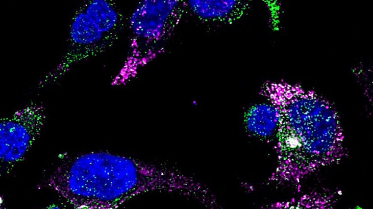
![GLP-1 and PYY localized to distinct secretory pools in L-cells. GLP-1 and PYY localized to distinct secretory pools in L-cells.](/fileadmin/_processed_/0/2/csm_L-cells_366ce08e69.jpg)
Cutting-Edge Imaging Techniques for GPCR Signaling
With this webinar on-demand enhance your pharmacological research with our webinar on GPCR signaling and explore cutting-edge imaging techniques that aim to understand how GPCR signaling translates…
Loading...
![Stripe assay performed on a THUNDER Imager Cell. Courtesy of Maria Carrasquero Ordaz, University of Oxford. Stripe assay performed on a THUNDER Imager Cell. Courtesy of Maria Carrasquero Ordaz, University of Oxford.](/fileadmin/_processed_/a/6/csm_Stripe_assay_THUNDER_Imager_Cell_429b2c2068.jpg)
Revealing Neuronal Migration’s Molecular Secrets
Different approaches can be used to investigate neuronal migration to their niche in the developing brain. In this webinar, experts from The University of Oxford present the microscopy tools and…
Loading...
![Dapi – Nucleus, GFP – Plasma Membrane, Thickness 100µm, 63x objektive, 469 Z planes, 2 channels, THUNDER Imager 3D Cell Culture. Courtesy M.Sc. Dana Krauß, Medical University of Vienna (Austria). Dapi – Nucleus, GFP – Plasma Membrane, Thickness 100µm, 63x objektive, 469 Z planes, 2 channels, THUNDER Imager 3D Cell Culture. Courtesy M.Sc. Dana Krauß, Medical University of Vienna (Austria).](/fileadmin/_processed_/5/a/csm_Two_color_organoid_cluster_afbae4f019.jpg)
How Efficient is your 3D Organoid Imaging and Analysis Workflow?
Organoid models have transformed life science research but optimizing image analysis protocols remains a key challenge. This webinar explores a streamlined workflow for organoid research, starting…