Werfen Sie einen Blick auf alle unsere anstehenden Konferenzen, Kongresse, Messen, Webinare und Workshops und besuchen Sie uns bei einer unserer nächsten Veranstaltungen!
22
–
23
Apr
2025
第四届半导体封装检测及失效分析技术进展网络研讨会
China
•
Webinar
22
Apr
2025
共聚焦荧光寿命成像功能在植物相关研究中的应用
China
•
Webinar
Filter articles
Tags
Produkte
Loading...
.")
How Efficient is your 3D Organoid Imaging and Analysis Workflow?
Organoid models have transformed life science research but optimizing image analysis protocols remains a key challenge. This webinar explores a streamlined workflow for organoid research, starting…
Loading...

Improve Your Ultramicrotomy Workflow with Automated Sectioning
Discover advanced digital ultramicrotomy tools for fast and accurate automated sectioning. Learn about autoalignment, and efficient sample trimming leveraging 3D µCT data. See application examples…
Loading...

How do Cells Talk to Each Other During Neurodevelopment?
Professor Silvia Capello presents her group’s research on cellular crosstalk in neurodevelopmental disorders, using models such as cerebral organoids and assembloids.
Loading...

Extended Live-cell Imaging at Nanoscale Resolution
Extended live-cell imaging with TauSTED Xtend. Combined spatial and lifetime information allow super-resolution microscopy at extremely low light dose.
Loading...
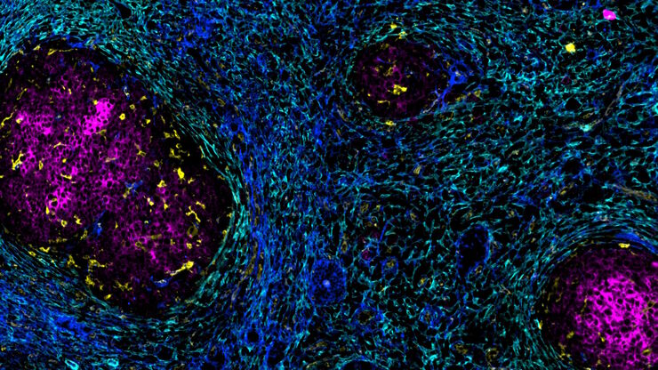
Accelerating Discovery for Multiplexed Imaging of Diverse Tissues
Explore IBEX: Open-source multiplexed imaging. Join the collaborative IBEX Imaging Community for optimized tissue processing, antibody selection, and human atlas construction.
Loading...

Transforming Multiplexed 2D Data into Spatial Insights Guided by AI
Aivia 13 handles large 2D images and enables researchers to obtain deep insights into microenvironment surrounding their phenotypes with millions of detected objects and automatic clustering up to 30…
