UC Enuity
Preparación de muestras de ME
Productos
Inicio
Leica Microsystems
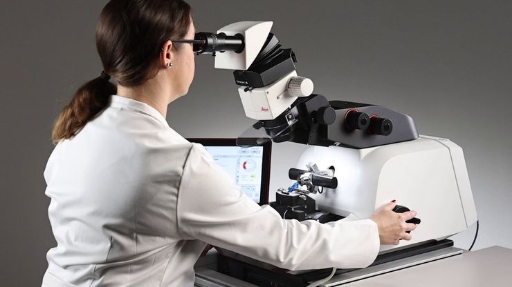
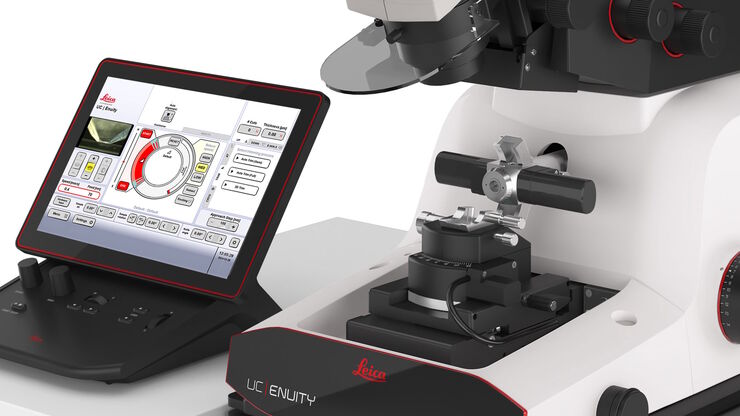
UC Enuity Ultramicrotomo
Experimente lo último en automatización y precisión
Lea nuestros últimos artículos
How to Save Time and Samples by Automated Ultramicrotomy
This article describes how 3D micro-CT data of a resin-embedded electron microscopy sample can be used to trim the specimen down to a defined target plane prior to sectioning. The interactive and…
Essential Guide to Ultramicrotomy
When studying samples, to visualize their fine structure with nanometer scale resolution, most often electron microscopy is used. There are 2 types: scanning electron microscopy (SEM) which images the…
How to Automatically Obtain Fluorescent Cells of Interest in a Block-face
Block-face created by automatic trimming under fluorescence.
Mammalian cells of interest, stained with CellTrackerTM Green are visualized within the block-face using the UC Enuity equipped with the…
Improve Your Ultramicrotomy Workflow with Automated Sectioning
Discover advanced digital ultramicrotomy tools for fast and accurate automated sectioning. Learn about autoalignment, and efficient sample trimming leveraging 3D µCT data. See application examples…
Workflow Solutions for Sample Preparation Methods for Material Science
This brochure presents and explains appropriate workflow solutions for the most frequently required sample preparation methods for material science samples.
Automatic Alignment of Sample and Knife for High Sectioning Quality
Automatic alignment of sample and knife on the ultramicrotome UC Enuity, enabling even untrained users to create ultrathin sections with reduced risk of losing precious sections.
High Quality Sectioning in Ultramicrotomy
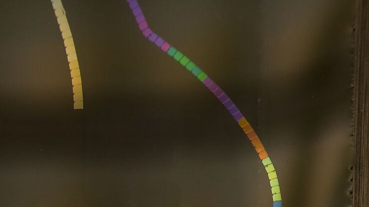
Discover the significance of achieving high-quality uniform sections with ultramicrotomy for precise imaging in electron microscopy.
Ultramicrotomy Techniques for Materials Sectioning
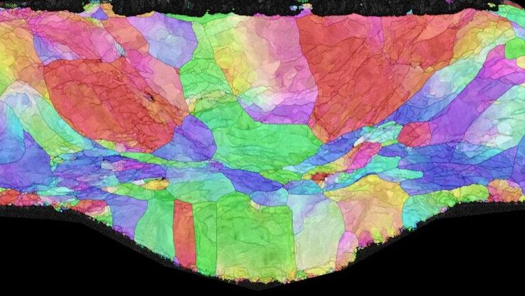
Learn about ultramicrotomy for materials sectioning when investigating polymers and brittle materials with transmission (TEM) or scanning electron microscopy (SEM) or atomic force microscopy.
Campos de aplicación
Microscopios para investigación en ciencias biológicas
Los microscopios de Leica Microsystems para investigación en ciencias biológicas satisfacen las necesidades de adquisición de imágenes de la comunidad científica gracias a un elevado grado de…
Microscopios para análisis de materiales
El análisis de materiales requiere microscopios para la obtención de imágenes, la medición y el análisis de características de diversos materiales, como aleaciones metálicas, semiconductores, vidrio y…


