
Science Lab
Science Lab
Bienvenido al portal de conocimiento de Leica Microsystems. Aquí encontrará investigación científica y material didáctico sobre el tema de la microscopía. El portal ayuda a principiantes, profesionales experimentados y científicos por igual en su trabajo diario y en sus experimentos. Explore tutoriales interactivos y notas de aplicación, descubra los fundamentos de la microscopía, así como las tecnologías de gama alta. Forme parte de la comunidad Science Lab y comparta sus conocimientos.
Filter articles
Etiquetas
Story Type
Products
Loading...

Overcoming Challenges with Microscopy when Imaging Moving Zebrafish Larvae
Zebrafish is a valuable model organism with many beneficial traits. However, imaging a full organism poses challenges as it is not stationary. Here, this case study shows how zebrafish larvae can be…
Loading...

Microscopios para disección
Las disecciones pueden implicar pasar horas mirando por los oculares de un microscopio para disección. Leica Microsystems le permite escoger entre una variada gama de microscopios y un amplio rango de…
Loading...

Neurociencia
¿Trabaja para comprender mejor las enfermedades neurodegenerativas o la función del sistema nervioso? Vea cómo puede conseguir descubrimientos con las soluciones de imágenes de Leica Microsystems.
Loading...

Mapping Tumor Immune Landscape with AI-Powered Spatial Proteomics
Spatial mapping of untreated tumors provides an overview of the tumor immune architecture, useful for understanding therapeutic responses. Immunocompetent murine models are essential for identifying…
Loading...

Virología
¿Su interés en investigación se centra en infecciones víricas y enfermedades? Descubra cómo puede adquirir conocimientos sobre virología con soluciones de generación de imágenes y preparación de…
Loading...
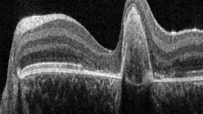
A Guide to OCT
Leica Optical Coherence Tomography (OCT) systems support ophthalmologists, ophthalmic surgeons, and researchers with easy-to-use, high-quality imaging technology.
Loading...

Tomografía Crioelectrónica
La tomografía crioelectrónica (CryoET) se utiliza para resolver biomoléculas dentro de su entorno celular hasta una resolución sin precedentes por debajo de un nanómetro.
Loading...

Investigación con Pez Cebra
Para obtener los mejores resultados en clasificación, selección, manipulación y captura y procesamiento de imágenes necesita ver los detalles y estructuras para poder tomar las decisiones adecuadas…
Loading...

Aneurysm Clipping: Assessing Perforators in Real-Time with AR Fluorescence
This article covers two aneurysm clipping cases highlighting the clinical benefits of GLOW800 Augmented Reality Fluorescence application in neurosurgery, based on insights from Prof. Tohru Mizutani,…
