
is mobile? false
Ciencias de la vida
Ciencias de la vida
Este es el lugar para ampliar sus conocimientos, capacidades de investigación y aplicaciones prácticas de la microscopía en diversos campos científicos. Aprenda a conseguir una visualización precisa, interpretación de imágenes y avances en la investigación. Encuentre información detallada sobre microscopía avanzada, técnicas de obtención de imágenes, preparación de muestras y análisis de imágenes. Los temas tratados incluyen la biología celular, la neurociencia y la investigación del cáncer, con especial atención a las aplicaciones e innovaciones de vanguardia.
Popular Show subnavigation
Ciencias de la vida


Filter articles
Etiquetas
Story Type
Products
Loading...

Virología
¿Su interés en investigación se centra en infecciones víricas y enfermedades? Descubra cómo puede adquirir conocimientos sobre virología con soluciones de generación de imágenes y preparación de…
Loading...

Epi-Illumination Fluorescence and Reflection-Contrast Microscopy
This article discusses the development of epi-illumination and reflection contrast for fluorescence microscopy concerning life-science applications. Much was done by the Ploem research group…
Loading...
, TL DIC")
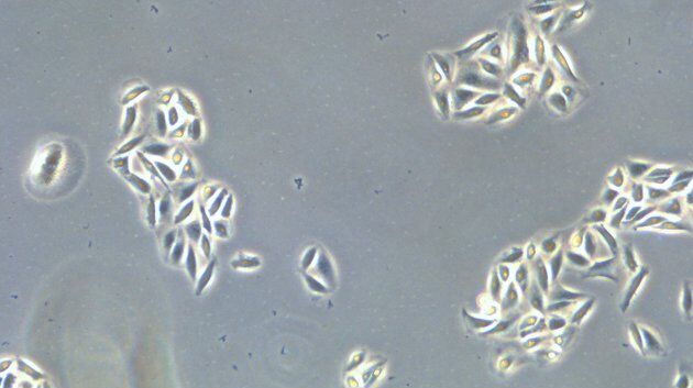
DIC
Un microscopio DIC es un microscopio de campo ancho que tiene un filtro de polarización y un prisma Wollaston entre la fuente de luz y la lente del condensador, así como entre la lente del objetivo y…
Loading...

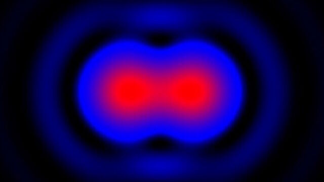
Microscopios de campo oscuro
El método de contraste de campo oscuro aprovecha la difracción o dispersión de la luz de las estructuras de un espécimen biológico o las características no uniformes de una muestra de material.
Loading...
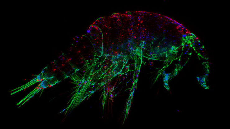
The Fundamentals and History of Fluorescence and Quantum Dots
At some point in your research and science career, you will no doubt come across fluorescence microscopy. This ubiquitous technique has transformed the way in which microscopists can image, tag and…
Loading...
 illuminated with wide-band UV excitation. Note the tissue structure with blue autofluorescence. Right: Same tissue and same immunostaining with FITC label illuminated with epi-illumination using narrow")
Milestones in Incident Light Fluorescence Microscopy
Since the middle of the last century, fluorescence microscopy developed into a bio scientific tool with one of the biggest impacts on our understanding of life. Watching cells and proteins with the…
Loading...

Video Tutorial: How to Change the Bulb of a Fluorescence Lamp Housing
When applying fluorescence microscopy in biological applications, a lamp housing with mercury burner is the most common light source. This video tutorial shows how to change the bulb of a traditional…
Loading...

Video Tutorial: How to Align the Bulb of a Fluorescence Lamp Housing
The traditional light source for fluorescence excitation is a fluorescence lamp housing with mercury burner. A prerequisite for achieving bright and homogeneous excitation is the correct centering and…
Loading...

Fluorescent Proteins - From the Beginnings to the Nobel Prize
Fluorescent proteins are the fundament of recent fluorescence microscopy and its modern applications. Their discovery and consequent development was one of the most exciting innovations for life…
