
Ciencias de la vida
Ciencias de la vida
Este es el lugar para ampliar sus conocimientos, capacidades de investigación y aplicaciones prácticas de la microscopía en diversos campos científicos. Aprenda a conseguir una visualización precisa, interpretación de imágenes y avances en la investigación. Encuentre información detallada sobre microscopía avanzada, técnicas de obtención de imágenes, preparación de muestras y análisis de imágenes. Los temas tratados incluyen la biología celular, la neurociencia y la investigación del cáncer, con especial atención a las aplicaciones e innovaciones de vanguardia.
Filter articles
Etiquetas
Story Type
Products
Loading...

From Bench to Beam: A Complete Correlative Cryo Light Microscopy Workflow
In the webinar entitled "A Multimodal Vitreous Crusade, a Cryo Correlative Workflow from Bench to Beam" a team of experts discusses the exciting world of correlative workflows for structural biology…
Loading...

Tomografía Crioelectrónica
La tomografía crioelectrónica (CryoET) se utiliza para resolver biomoléculas dentro de su entorno celular hasta una resolución sin precedentes por debajo de un nanómetro.
Loading...
Advancing Cellular Ultrastructure Research
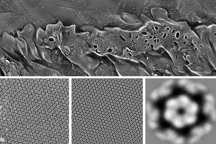
Freeze-fracture and freeze-etching are useful tools for studying flexible membrane-associated structures such as tight junctions or the enteric glycocalyx. Freeze-fracture and etching are two…
Loading...

Exploring the Structure and Life Cycle of Viruses
The SARS-CoV-2 outbreak started in late December 2019 and has since reached a global pandemic, leading to a worldwide battle against COVID-19. The ever-evolving electron microscopy methods offer a…
Loading...

Workflows and Instrumentation for Cryo-electron Microscopy
Cryo-electron microscopy is an increasingly popular modality to study the structures of macromolecular complexes and has enabled numerous new insights in cell biology. In recent years, cryo-electron…
Loading...

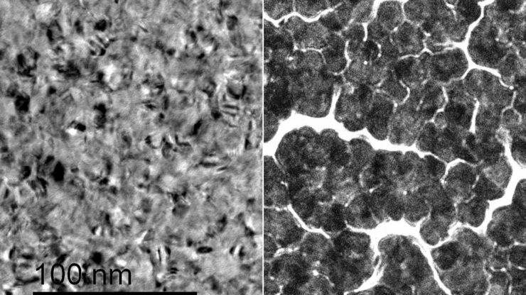
Macroscale to Nanoscale Pore Analysis of Shale and Carbonate Rocks
Physical porosity in rocks, like shale and carbonate, has a large effect on the their storage capacity. The pore geometries also affect their permeability. Imaging the visible pore space provides…
Loading...

Visualization of DNA Molecules
Precise low angle rotary shadowing with heavy metals (like platinum) can be used in transmission electron microscopy (TEM) to observe molecular details of objects previously absorbed on a thin, low…
Loading...
Improvement of Metallic Thin Films for HR-SEM by Using DC Magnetron Sputter Coater
Preparation techniques, like several kinds of coating methods play an important role for high resolution scanning electron microscopy (HR-SEM). Nonconductive sample like biological and synthetic…
Loading...
Brief Introduction to Freeze Fracture and Etching
Freeze fracture describes the technique of breaking a frozen specimen to reveal internal structures. Freeze etching is the sublimation of surface ice under vacuum to reveal details of the fractured…
