
Ciencias de la vida
Ciencias de la vida
Este es el lugar para ampliar sus conocimientos, capacidades de investigación y aplicaciones prácticas de la microscopía en diversos campos científicos. Aprenda a conseguir una visualización precisa, interpretación de imágenes y avances en la investigación. Encuentre información detallada sobre microscopía avanzada, técnicas de obtención de imágenes, preparación de muestras y análisis de imágenes. Los temas tratados incluyen la biología celular, la neurociencia y la investigación del cáncer, con especial atención a las aplicaciones e innovaciones de vanguardia.
Filter articles
Etiquetas
Story Type
Products
Loading...
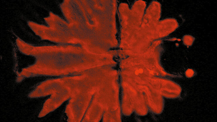
 applied. Image courtesy of Samuel East, Uncommon Bio.")
Designing the Future with Novel and Scalable Stem Cell Culture
Visionary biotech start-up Uncommon Bio is tackling one of the world’s biggest health challenges: food sustainability. In this webinar, Stem Cell Scientist Samuel East shows how they make stem cell…
Loading...
A Guide to Phase Contrast
A phase contrast light microscope offers a way to view the structures of many types of biological specimens in greater contrast without the need of stains.
Loading...

Neurociencia
¿Trabaja para comprender mejor las enfermedades neurodegenerativas o la función del sistema nervioso? Vea cómo puede conseguir descubrimientos con las soluciones de imágenes de Leica Microsystems.
Loading...

Virología
¿Su interés en investigación se centra en infecciones víricas y enfermedades? Descubra cómo puede adquirir conocimientos sobre virología con soluciones de generación de imágenes y preparación de…
Loading...

Investigación con Pez Cebra
Para obtener los mejores resultados en clasificación, selección, manipulación y captura y procesamiento de imágenes necesita ver los detalles y estructuras para poder tomar las decisiones adecuadas…
Loading...

Revealing Neuronal Migration’s Molecular Secrets
Different approaches can be used to investigate neuronal migration to their niche in the developing brain. In this webinar, experts from The University of Oxford present the microscopy tools and…
Loading...
.")
How Efficient is your 3D Organoid Imaging and Analysis Workflow?
Organoid models have transformed life science research but optimizing image analysis protocols remains a key challenge. This webinar explores a streamlined workflow for organoid research, starting…
Loading...
 where cyan indicates nuclei and magenta tight junctions.")
Rapid Check of Live Stem Cells in Cell-Culture Inserts set in Multi-Well Plates
See how efficient imaging of live iPSC stem cells within cell-culture inserts set in a multi-well plate can be done to evaluate the cells using a THUNDER Imager. Just read this article.
Loading...
 imaged with the THUNDER Imager 3D Cell Culture. Courtesy of Dr. F.T. Arroso Martins, Tamere University, Finland.")
How to Get Deeper Insights into your Organoid and Spheroid Models
In this eBook, learn about key considerations for imaging 3D cultures, such as organoids and spheroids, and discover microscopy solutions to shed new insights into dynamic processes in 3D real-time
