
Science Lab
Science Lab
Bienvenido al portal de conocimiento de Leica Microsystems. Aquí encontrará investigación científica y material didáctico sobre el tema de la microscopía. El portal ayuda a principiantes, profesionales experimentados y científicos por igual en su trabajo diario y en sus experimentos. Explore tutoriales interactivos y notas de aplicación, descubra los fundamentos de la microscopía, así como las tecnologías de gama alta. Forme parte de la comunidad Science Lab y comparta sus conocimientos.
Filter articles
Etiquetas
Story Type
Products
Loading...

Optimizing THUNDER Platform for High-Content Slide Scanning
With rising demand for full-tissue imaging and the need for FL signal quantitation in diverse biological specimens, the limits on HC imaging technology are tested, while user trainability and…
Loading...

Physiology Image Gallery
Physiology is about the processes and functions within a living organism. Research in physiology focuses on the activities and functions of an organism’s organs, tissues, or cells, including the…
Loading...
, TL DIC")
DIC
Un microscopio DIC es un microscopio de campo ancho que tiene un filtro de polarización y un prisma Wollaston entre la fuente de luz y la lente del condensador, así como entre la lente del objetivo y…
Loading...

Microscopios de campo oscuro
El método de contraste de campo oscuro aprovecha la difracción o dispersión de la luz de las estructuras de un espécimen biológico o las características no uniformes de una muestra de material.
Loading...

Developmental Biology Image Gallery
Developmental biology explores the development of complex organisms from the embryo to adulthood to understand in detail the origins of disease. This category of the gallery shows images about…
Loading...

The Power of Pairing Adaptive Deconvolution with Computational Clearing
Learn how deconvolution allows you to overcome losses in image resolution and contrast in widefield fluorescence microscopy due to the wave nature of light and the diffraction of light by optical…
Loading...
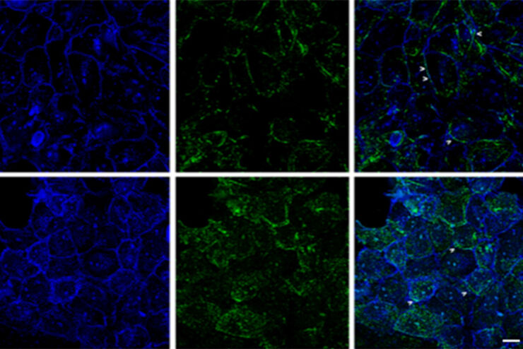
Improvement of Imaging Techniques to Understand Organelle Membrane Cell Dynamics
Understanding cell functions in normal and tumorous tissue is a key factor in advancing research of potential treatment strategies and understanding why some treatments might fail. Single-cell…
Loading...

Image Gallery: THUNDER Imager
To help you answer important scientific questions, THUNDER Imagers eliminate the out-of-focus blur that clouds the view of thick samples when using camera-based fluorescence microscopes. They achieve…
Loading...
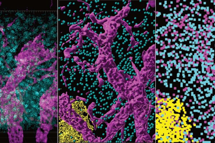
From Organs to Tissues to Cells: Analyzing 3D Specimens with Widefield Microscopy
Obtaining high-quality data and images from thick 3D samples is challenging using traditional widefield microscopy because of the contribution of out-of-focus light. In this webinar, Falco Krüger…
