Filter articles
Etiquetas
Products
Loading...

From Bench to Beam: A Complete Correlative Cryo Light Microscopy Workflow
In the webinar entitled "A Multimodal Vitreous Crusade, a Cryo Correlative Workflow from Bench to Beam" a team of experts discusses the exciting world of correlative workflows for structural biology…
Loading...
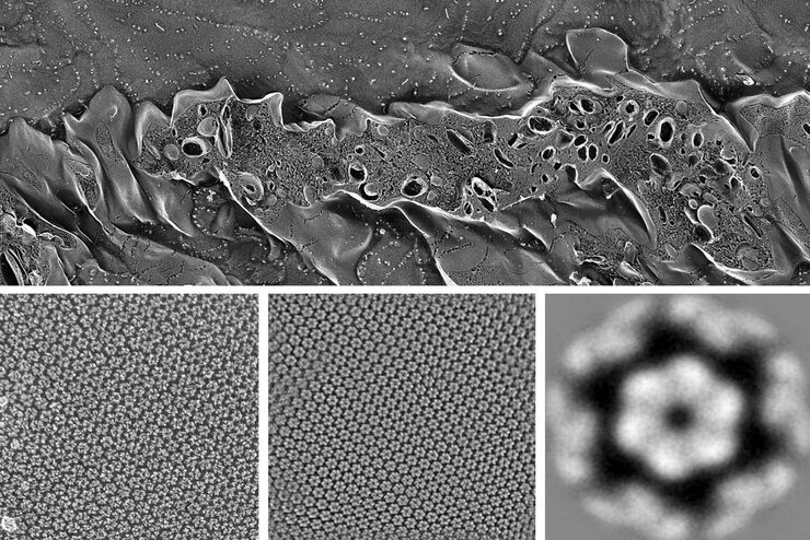
Advancing Cellular Ultrastructure Research
Freeze-fracture and freeze-etching are useful tools for studying flexible membrane-associated structures such as tight junctions or the enteric glycocalyx. Freeze-fracture and etching are two…
Loading...

Exploring the Structure and Life Cycle of Viruses
The SARS-CoV-2 outbreak started in late December 2019 and has since reached a global pandemic, leading to a worldwide battle against COVID-19. The ever-evolving electron microscopy methods offer a…
Loading...

Workflows and Instrumentation for Cryo-electron Microscopy
Cryo-electron microscopy is an increasingly popular modality to study the structures of macromolecular complexes and has enabled numerous new insights in cell biology. In recent years, cryo-electron…
