
Science Lab
Science Lab
Bienvenido al portal de conocimiento de Leica Microsystems. Aquí encontrará investigación científica y material didáctico sobre el tema de la microscopía. El portal ayuda a principiantes, profesionales experimentados y científicos por igual en su trabajo diario y en sus experimentos. Explore tutoriales interactivos y notas de aplicación, descubra los fundamentos de la microscopía, así como las tecnologías de gama alta. Forme parte de la comunidad Science Lab y comparta sus conocimientos.
Filter articles
Etiquetas
Story Type
Products
Loading...

Consumables for Laser Microdissection
There are many different types of consumables for laser microdissection (LMD) systems. They cover a wide range of applications from basic to highly specialized, enabling scientists to choose their own…
Loading...
Introduction to Ion Beam Etching with the EM TIC 3X
In this article you can learn how to optimize the preparation quality of your samples by using the ion beam etching method with the EM TIC 3X ion beam milling machine. A short introduction of the…
Loading...

How to Drape an Overhead Surgical Microscope
The tutorial features the Leica ARveo digital Augmented Reality microscope for complex neurosurgery. The procedure also applies to the Leica M530 OHX, OH6, OH5 and OH4.
Loading...

How to Drape a Surgical Microscope
Before performing surgical procedures, it is important to drape the surgical microscope to ensure sterile working conditions. At Leica, we are committed to helping you with your surgical practice. In…
Loading...
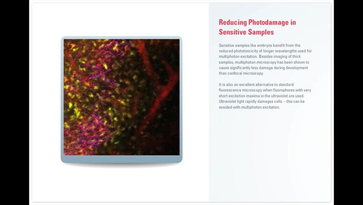
Principles of Multiphoton Microscopy for Deep Tissue Imaging
This tutorial explains the principles of multiphoton microscopy for deep tissue imaging. Multiphoton microscopy uses excitation wavelengths in the infrared taking advantage of the reduced scattering…
Loading...
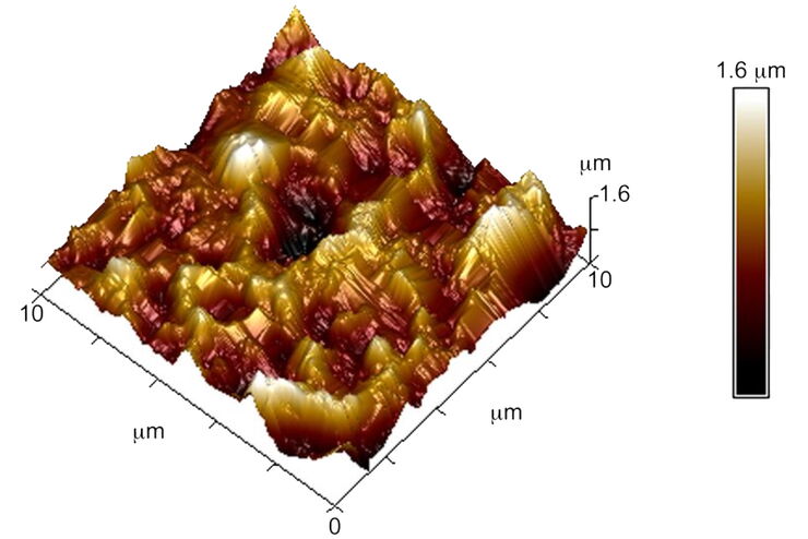
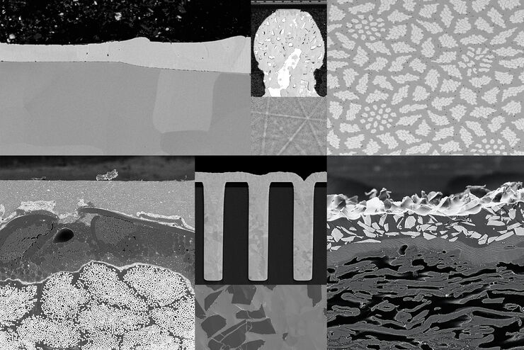
Brief Introduction to Surface Metrology
This report briefly discusses several important metrology techniques and standard definitions commonly used to assess the topography of surfaces, also known as surface texture or surface finish. With…
Loading...
 images")
How To Create EDOF (Extended Depth of Focus) Images
Watch this video to see how you can rapidly record sharp optical microscope images of samples with a large height variation. This is done with the optional Extended Depth of Focus (EDOF) function of…
Loading...
![3D glomeruli in a portion of an ECi-cleared kidney scanned by light sheet microscopy. Courtesy of Prof. Norbert Gretz, Medical Faculty Mannheim, University of Heidelberg [1].](/fileadmin/_processed_/d/d/csm_DLS-Sample-Preparation-Intr_915e0fd7c2.jpg "3D glomeruli in a portion of an ECi-cleared kidney scanned by light sheet microscopy. Courtesy of Prof. Norbert Gretz, Medical Faculty Mannheim, University of Heidelberg [1].")
Using Mounting Frames for Light Sheet Sample Preparation
Sample handling is an important topic in the context of Light Sheet Microscopy. The TCS SP8 DLS integrates Light Sheet technology into an inverted confocal platform and can hence make use of general…
Loading...

Using a Rotation Device for Light Sheet Sample Mounting
The TCS SP8 DLS from Leica Microsystems is an innovative concept to integrate the Light Sheet Microscopy technology into the confocal microscope. Due to its unique optical architecture samples can be…
