Proveo 8
Microscope opératoire
Produits
Accueil
Leica Microsystems
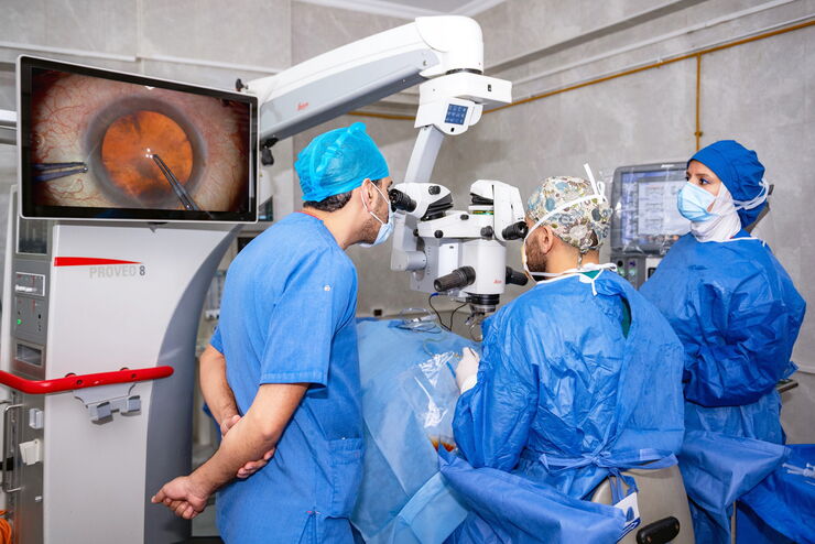
Proveo 8 Microscope ophtalmique
Une efficacité perceptible, une précision fiable
Lire nos derniers articles
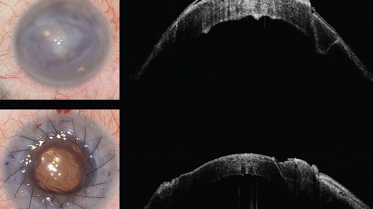
How does Real-Time OCT Imaging Impact Precision in Corneal Surgery?
Corneal surgery is a highly specialized field. It requires great surgical precision to overcome challenges such as visualizing clearly the full anterior chamber, performing Descemet membrane peeling…
RPE65 Gene Therapy Subretinal Injection: Benefits of Intraoperative OCT
Discover how RPE65 gene therapy subretinal injection procedures in patients with Leber congenital amaurosis is supported by intraoperative Optical Coherence Tomography.
Dislocated Cataract Angle Closure Aided by Intraoperative OCT
Learn how a dislocated cataract was treated with angle closure assisted by intraoperative OCT to achieve long-term good results without future lens dislocation.
Glaucoma Stent Revision Surgery Guided by Intraoperative OCT
Learn about a glaucoma subconjunctival stent revision guided by intraoperative OCT and the important role it plays to ensure the best outcome.
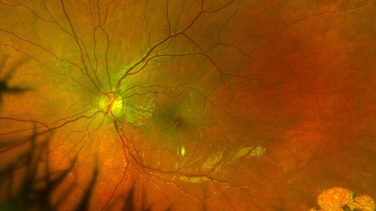
Posterior Segment Surgery: Benefits of Utilizing Intraoperative OCT
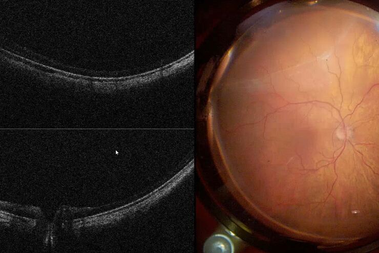
Learn about the value of intraoperative optical coherence tomography in posterior segment surgery to precisely locate, evaluate and manage pathologies.
Intraoperative OCT-Assisted Corneal Transplant Procedures
Learn about the use of intraoperative optical coherence tomography in corneal transplantation and how it facilitates the adaptation of the donor cornea.
Improve Macular Hole Surgery with Optical Coherence Tomography
A case study on the use of intraoperative OCT during macular hole surgery for pediatric lamellar macular hole repair and how it provides valuable real-time information.
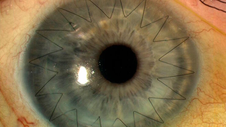
Ophthalmology Case Study: Corneal Transplantation
Learn about the use of intraoperative Optical Coherence Tomography in Corneal Transplantation and how it helps achieve correct positioning of donor tissue.
Utility of Intraoperative OCT in Sub-Retinal Gene Therapy
Discover a case study on the use of intraoperative OCT for pediatric gene therapy and how it supports bleb placement and verifying the foveal contour.
Ophthalmic Gene Therapy Subretinal Injection
Case study on the use of intraoperative OCT for Leber congenital amaurosis macular repair and ophthalmic gene therapy subretinal injection.
Dr. Tawfik Shares his Expert View on Direct Horizontal Chopping in Cataract Surgery
It is estimated that nearly 28 million cataract surgery procedures are performed worldwide every year. Phacoemulsification is the most common method used to remove the cataract and chopping maneuvers…
Clinical Symposium on OCT-Guided Retina Surgery
In this recording Prof. Tan from Singapore National Eye Centre and Dr. Català from Sant Joan de Déu Barcelona Children’s Hospital share their expertise on retinal surgery with intraoperative OCT from…
How to Select a Microscope for Cataract Surgery
What to consider in the selection of an ophthalmic microscope for cataract procedures. Bearing these aspects in mind will equip surgeons well for talks with manufacturer representatives. Many…
Proveo 8 with intraoperative OCT – a User Evaluation in an University Setting
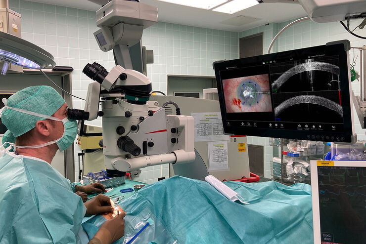
Optical coherence tomography (OCT) makes structures in the eye visible that lie beneath the surface. When OCT is used intraoperatively, surgeons gain insight into possible pathological changes in the…
Intraoperative OCT in Retinal Procedures
In their white paper the surgical team at Retina Clinic in São Paulo provide insight into cases of complex retinal surgery in which OCT has provided helpful additional information which has on…
Clinical Symposium on OCT-guided Cornea Surgery
In this recording Prof. Mehta from Singapore National Eye Centre and Prof. Fontana from Santa Maria
Nuova Hospital in Regio Emilia, Italy, share their expertise on corneal surgery. They present PK,…
Intraoperative OCT-Assisted Surgical Management of Proliferative Vitreoretinopathy
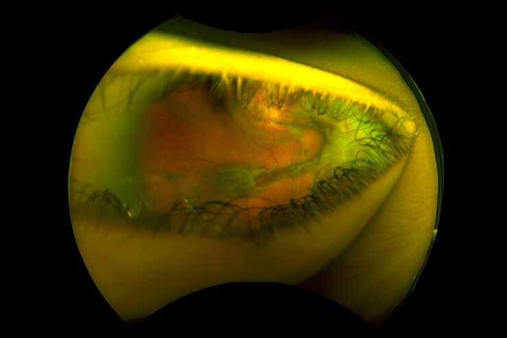
Proliferative vitreoretinopathy (PVR) is a plague to patients and their surgeons after recent rhegmatogenous retinal detachment (RD). Despite excellent initial surgical outcomes, it is the most common…
Overcoming Ophthalmologic Surgery Challenges
Ophthalmology surgical procedures involving both the anterior and posterior segment can be particularly challenging. Good visualization is required to operate with precision and confidence.
Prof.…
Moving to Routine Use of Intraoperative OCT in Eye Surgery
Dr Barbara Parolini is one of the pioneers in the use of intraoperative OCT in eye surgery. Since 2016, she has gained a lot of clinical experience with different intraoperative OCT systems. In this…
How OCT-guided Eye Surgery can help you to Focus on Perfection
Watch the successful clinical online symposium on-demand. Find out what Dr. Parolini says about the benefits of OCT-guided ophthalmic surgery. She presents clinical cases and shares her experience…
Advanced Techniques in Cataract and Refractive Surgery
In this webinar Dr. Thompson and Dr. Moshirfar will explain how Leica microscopes aid in procedures such as Centration of Multifocal IOLs and corneal inlays such as Kamra and Lenticular Grafts used in…
Cataract Surgery with CoAx4 Illumination
A stable red reflex is one of the most important features of an ophthalmic surgical microscope for cataract surgery. It’s the red reflex that makes the structure of the lens visible and thus makes for…
FusionOptics in Neurosurgery and Ophthalmology – for a Larger 3D Area in Focus
Neurosurgeons and ophthalmologists deal with delicate structures, deep or narow cavities and tiny structures with vitally important functions. A clear, three-dimensional view on the surgical field is…
Domaines d'application
Ophtalmologie
Les microscopes opératoires ophtalmologiques de Leica excellent dans le domaine de l'optique et de l'éclairage, aidant les chirurgiens à réaliser des chirurgies avec une précision supérieure et à…
Chirurgie de la cataracte
Les opérations de la cataracte sont devenues l'une des procédures de chirurgie ophtalmiques les plus fréquentes au cours des dernières décennies. Le chirurgien ophtalmologiste retire la cataracte et…
Chirurgie de la rétine
Vous recherchez des solutions de microscopie pour la chirurgie de la rétine ? Découvrez les avantages uniques des solutions Leica et comment elles peuvent vous aider à relever vos défis.
Chirurgie de la cornée
Les microscopes ophtalmiques Leica offrent de nombreuses solutions pour la chirurgie de la cornée et vous assistent depuis le prélèvement et la préparation des tissus cornéens du donneur jusqu'à la…
Chirurgie du glaucome
Les solutions de microscopie ophtalmique Leica offrent une vision optimale pour la trabéculectomie, la chirurgie du glaucome par implant et la chirurgie mini-invasive du glaucome (MIGS).


