STELLARIS STED
Microscopes confocaux
Produits
Accueil
Leica Microsystems
STELLARIS STED
Des données fiables, plus vite !
Lire nos derniers articles
, and trafficking vesicles labeled with CF594 (cyan - Biotium).")
A Guide to Super-Resolution
Find out more about Leica super-resolution microscopy solutions and how they can empower you to visualize in fine detail subcellular structures and dynamics.

Super-Resolution Microscopy Image Gallery
Due to the diffraction limit of light, traditional confocal microscopy cannot resolve structures below ~240 nm. Super-resolution microscopy techniques, such as STED, PALM or STORM or some…

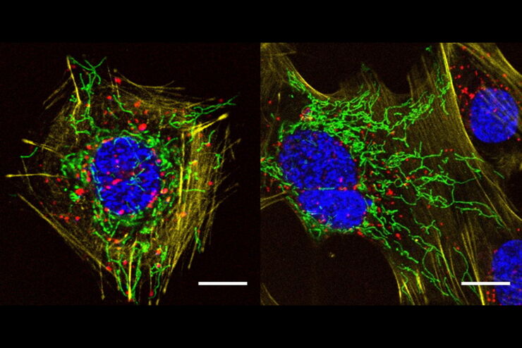
Extended Live-cell Imaging at Nanoscale Resolution
Extended live-cell imaging with TauSTED Xtend. Combined spatial and lifetime information allow super-resolution microscopy at extremely low light dose.

Studying Virus Replication with Fluorescence Microscopy
The results from research on SARS-CoV-2 virus replication kinetics, adaption capabilities, and cytopathology in Vero E6 cells, done with the help of fluorescence microscopy, are described in this…

Five-color FLIM-STED with One Depletion Laser
Webinar on five-color STED with a single depletion laser and fluorescence lifetime phasor separation.
A Versatile Palette of Fluorescent Probes
Researchers at the Max Planck Institute for Medical Research in Heidelberg have developed a general strategy to synthesize live-cell compatible fluorogenic probes, and the result are the new MaP (Max…

Benefits of Combining STED and Lifetime
In this interview, Professor Alberto Diaspro talks about the advantages of the White Light Laser and the TauSTED capabilities of STELLARIS 8 STED. He speaks about his experience with the confocal…
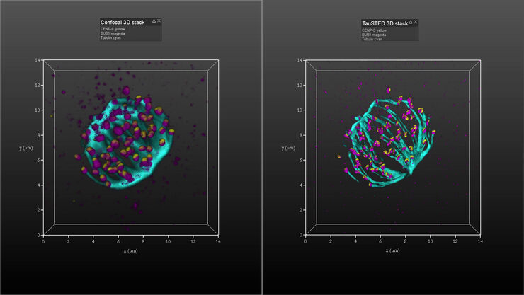
Kinetochore Assembly during Mitosis with TauSTED on 3D
Three-dimensional organization of the mitotic spindle together with the distribution of CENP-C and BUB1 based on TauSTED with multiple STED lines (592, 660 and 775 nm) can provide insights…

Regulators of Actin Cytoskeletal Regulation and Cell Migration in Human NK Cells
Dr. Mace will describe new advances in our understanding of the regulation of human NK cell actin cytoskeletal remodeling in cell migration and immune synapse formation derived from confocal and…
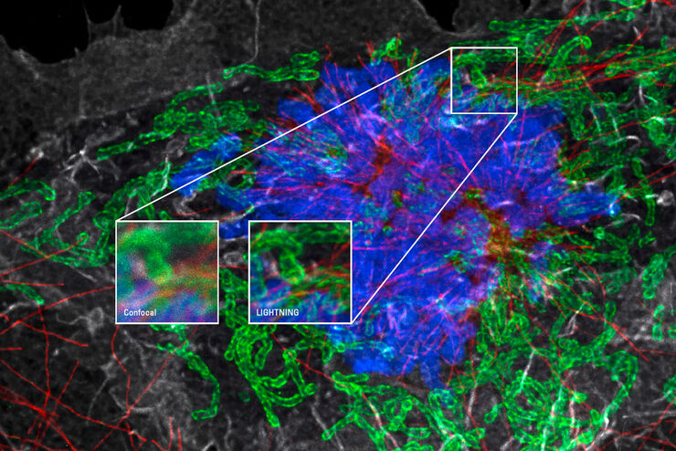
Obtenez le maximum d’informations à partir de votre échantillon grâce à LIGHTNING
LIGHTNING est un processus adaptatif d’extraction d’informations qui révèle les structures fines et les détails, parfois invisibles, et ce automatiquement. Contrairement aux technologies…

Universal PAINT – Dynamic Super-Resolution Microscopy
Super-resolution microscopy techniques have revolutionized biology for the last ten years. With their help cellular components can now be visualized at the size of a protein. Nevertheless, imaging…
Super-Resolution GSDIM Microscopy
The nanoscopic technique GSDIM (ground state depletion microscopy followed by individual molecule return) provides a detailed image of the spatial arrangement of proteins and other biomolecules within…
