
Science Lab
Science Lab
Bienvenue sur le portail de connaissances de Leica Microsystems. Vous y trouverez des recherches scientifiques et du matériel didactique sur le thème de la microscopie. Le portail aide les débutants, les praticiens expérimentés et les scientifiques dans leur travail quotidien et leurs expériences. Explorez les didacticiels interactifs et les notes d'application, découvrez les bases de la microscopie ainsi que les technologies de pointe. Faites partie de la communauté Science Lab et partagez votre expertise.
Filter articles
Tags
Story Type
Products
Loading...

Overcoming Challenges with Microscopy when Imaging Moving Zebrafish Larvae
Zebrafish is a valuable model organism with many beneficial traits. However, imaging a full organism poses challenges as it is not stationary. Here, this case study shows how zebrafish larvae can be…
Loading...

Microscopes de dissection
Si vous devez procéder à des dissections, il peut vous arriver de rester penché sur les oculaires du microscope pendant des heures. Leica Microsystems vous propose de faire votre choix parmi un large…
Loading...

Neurosciences
Vous souhaitez une meilleure compréhension des maladies neurodégénératives ou vous étudiez le fonctionnement du système nerveux ? Découvrez comment vous pouvez faire des percées importantes grâce aux…
Loading...

Mapping Tumor Immune Landscape with AI-Powered Spatial Proteomics
Spatial mapping of untreated tumors provides an overview of the tumor immune architecture, useful for understanding therapeutic responses. Immunocompetent murine models are essential for identifying…
Loading...

Virologie
Vos recherches portent sur les infections et les maladies virales ? Découvrez comment renforcer vos connaissances en virologie grâce aux solutions d’imagerie et de préparation d’échantillons de Leica…
Loading...
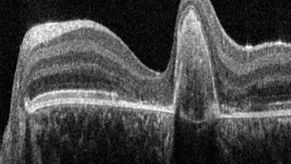
A Guide to OCT
Leica Optical Coherence Tomography (OCT) systems support ophthalmologists, ophthalmic surgeons, and researchers with easy-to-use, high-quality imaging technology.
Loading...

Tomographie Cryoélectronique
La tomographie cryoélectronique (CryoET) est utilisée pour analyser les biomolécules dans leur environnement cellulaire avec une résolution sans précédent, inférieure au nanomètre.
Loading...

A Guide to Zebrafish Research
For the best result during screening, sorting, manipulation, and imaging you need to see details and structures to make the right decisions for your next steps in research.
Known for outstanding…
Loading...

Aneurysm Clipping: Assessing Perforators in Real-Time with AR Fluorescence
This article covers two aneurysm clipping cases highlighting the clinical benefits of GLOW800 Augmented Reality Fluorescence application in neurosurgery, based on insights from Prof. Tohru Mizutani,…
