
Sciences de la vie
Sciences de la vie
C'est ici que vous pourrez développer vos connaissances, vos capacités de recherche et les applications pratiques de la microscopie dans divers domaines scientifiques. Apprenez à obtenir une visualisation précise, à interpréter les images et à faire progresser la recherche. Trouvez des informations pertinentes sur la microscopie avancée, les techniques d'imagerie, la préparation des échantillons et l'analyse des images. Les sujets abordés comprennent la biologie cellulaire, les neurosciences et la recherche sur le cancer, en mettant l'accent sur les applications et les innovations de pointe.
Filter articles
Tags
Story Type
Products
Loading...

Studying Wound Healing of Smooth Muscle Cells
This article discusses how wound healing of cultured smooth muscle cells (SMCs) in multiwell plates can be reliably studied over time with less effort using a specially configured Leica inverted…
Loading...
A Versatile Palette of Fluorescent Probes
Researchers at the Max Planck Institute for Medical Research in Heidelberg have developed a general strategy to synthesize live-cell compatible fluorogenic probes, and the result are the new MaP (Max…
Loading...

Formulated Product Characterization with SRS Microscopy
Creams, pastes, gels, emulsions, and tablets are ubiquitous across a wide range of manufacturing sectors from pharmaceuticals and consumer health products to agrochemicals and paint. To improve…
Loading...

Spectroscopic Evaluation of Red Blood Cells
Hemoglobinopathies are a major healthcare problem. This study presents a possible diagnostic tool for thalassemia which is based on confocal spectroscopy. This approach exploits spectral detection and…
Loading...
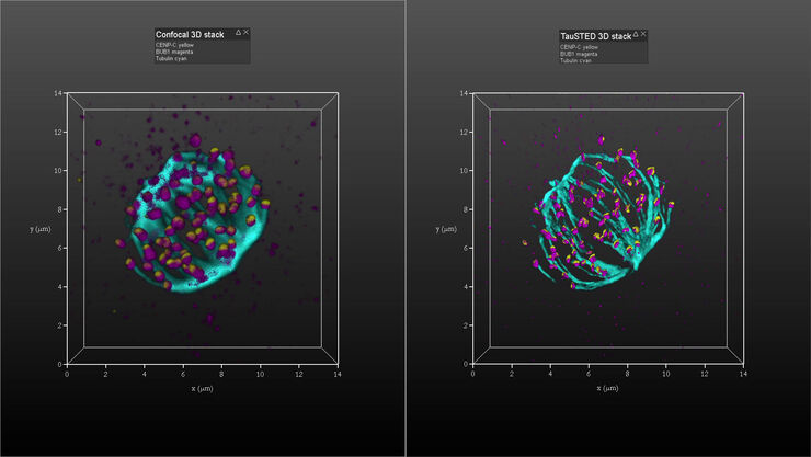
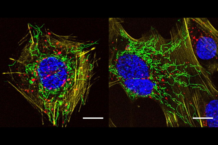
Kinetochore Assembly during Mitosis with TauSTED on 3D
Three-dimensional organization of the mitotic spindle together with the distribution of CENP-C and BUB1 based on TauSTED with multiple STED lines (592, 660 and 775 nm) can provide insights…
Loading...
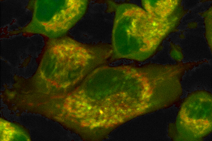
How to Quantify Changes in the Metabolic Status of Single Cells
Metabolic imaging based on fluorescence lifetime provides insights into the metabolic dynamics of cells, but its use has been limited as expertise in advanced microscopy techniques was needed.
Now,…
Loading...
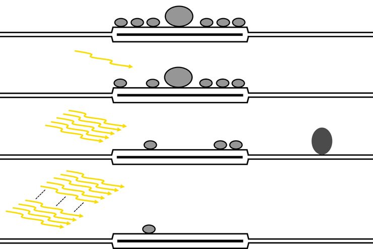
Investigating Synapses in Brain Slices with Enhanced Functional Electron Microscopy
A fundamental question of neuroscience is: what is the relationship between structural and functional properties of synapses? Over the last few decades, electrophysiology has shed light on synaptic…
Loading...

How FLIM Microscopy Helps to Detect Microplastic Pollution
The use of autofluorescence in biological samples is a widely used method to gain detailed knowledge about systems or organisms. This property is not only found in biological systems, but also…
