is mobile? false
Prodotti
Prodotti
Sviluppiamo i nostri sistemi di precisione high-tech per l'analisi delle microstrutture con l'utente, per l'utente. Il nostro portfolio prodotti include soluzioni di sistema destinate all'ambito delle Life Sciences, comprese biotecnologie e medicina, come pure soluzioni per la ricerca e lo sviluppo di materie prime e il controllo di qualità industriale.
Product Categories Show subnavigation
Prodotti
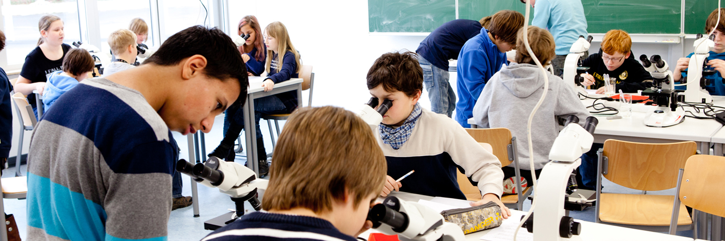
Prodotti per microscopio di precisione per l' analisi di microstrutture. Soluzioni per microscopio per scienza della vita, medicina, R&S e garanzia di qualità industriale.


Ultime Novità Show subnavigation
Software di analisi delle immagini
Aivia. Il futuro della microscopia basata sull'IA

Il pluripremiato microscopio d'ispezione
L'Emspira 3, che integra confronto, misurazione e condivisione della documentazione in un unico sistema, ha vinto un prestigioso Red Dot Award.
Eventi Show subnavigation
Date un'occhiata a tutte le nostre prossime conferenze, congressi, fiere, webinar e workshop e unitevi a noi in uno dei nostri prossimi eventi!
Applicazioni Show subnavigation


Leica Science Lab Articles Show subnavigation
Leggi gli articoli più recenti
Il portale informativo di Leica Microsystems offre materiale didattico e di ricerca scientifica su vari temi della microscopia. Il contenuto è stato progettato per aiutare i principianti, i professionisti esperti e gli scienziati nel lavoro quotidiano e negli esperimenti.
Essential Guide to Ultramicrotomy
When studying samples, to visualize their fine structure with nanometer scale resolution, most often electron microscopy is used. There are 2 types: scanning electron microscopy (SEM) which images the sample surface or transmission electron microscopy (TEM) which requires a very thin, electron-transparent sample. Thus, to image the fine structure inside a sample using electron microscopy, the solution is to make very thin sections of it.

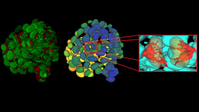
Explore Alzheimer's Spatial Proteome with Big Data
Alzheimer's disease, a genetic and sporadic neurodegenerative condition, leads to cognitive decline in mid to late life, marked by β-amyloid plaques and tau tangles. With limited treatment options, new investigative strategies are crucial. The Cell DIVE multiplexed imaging solution allows examination of Alzheimer's brain tissue, potentially uncovering new research avenues. Here, we showcase the Cell DIVE image viewer, enabling users to access the full Alzheimer's multiplexed dataset directly in their browser.
Ricerca sul cancro
Il cancro è una malattia complessa ed eterogenea causata da cellule che hanno dei difetti nella regolazione della crescita. I cambiamenti genetici ed epigenetici in una cellula o in un gruppo di cellule alterano il normale funzionamento e provocano una crescita e una proliferazione cellulare autonoma e incontrollata.

Dive into Pancreatic Cancer Research with Big Data
Pancreatic cancer, with a mortality rate near 40%, is challenging to treat due to its proximity to major organs. This story explores the complex biology of pancreatic ductal adenocarcinoma (PDAC), examining molecular and spatial determinants of tumor aggression in metabolism, apoptosis, and immunity. Access the full Cell DIVE dataset in your browser to delve deeper into these findings.

Uncover the Hidden Complexity of Colon Cancer with Big Data
Colorectal cancer poses a significant health burden. While surgery is effective initially, some patients develop recurrent secondary disease with poor prognosis, necessitating advanced therapies like immunotherapies. Spatial biology approaches, such as multiplexed imaging with Cell DIVE, can provide crucial insights for developing novel treatments. Access the full Cell DIVE dataset in your browser to explore these findings further through the Minerva image viewer.

Introduction to 21 CFR Part 11 for Electronic Records of Cell Culture
This article provides an introduction to the recommendations of 21 CFR Part 11 from the FDA, specifically focusing on the audit trail and user management in the context of cell-culture laboratories. It is intended for professionals in the biotechnology and pharmaceutical industries who are responsible for ensuring agreement with 21 CFR part 11 for electronic records and electronic signatures. A digital microscope approach, e.g. with Mateo FL, offers the advantage of more consistent and efficient electronic documentation of cell-culture results compared to a paper-based method.
Contrasto di fase
Grazie al microscopio ottico a contrasto di fase è possibile visualizzare le strutture di molti tipi di campioni biologici con maggior contrasto senza la necessità di utilizzare le colorazioni.
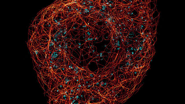
A Guide to Super-Resolution
Find out more about Leica super-resolution microscopy solutions and how they can empower you to visualize in fine detail subcellular structures and dynamics.
