
Specialità mediche
Specialità mediche
Esplora una raccolta completa di risorse scientifiche e cliniche su misura per gli operatori sanitari, tra cui approfondimenti tra pari, casi clinici e simposi. Progettata per neurochirurghi, oculisti e specialisti in chirurgia plastica e ricostruttiva, otorinolaringoiatria e odontoiatria. Questa raccolta evidenzia gli ultimi progressi nel campo della microscopia chirurgica. Scoprite come le tecnologie chirurgiche all'avanguardia, come la fluorescenza AR, la visualizzazione 3D e l'imaging OCT intraoperatorio, consentano di prendere decisioni sicure e di essere precisi in interventi chirurgici complessi.
Filter articles
Tag
Products
Loading...
Harnessing Microfluidics to Maintain Cell Health During Live-Cell Imaging
VIDEO ON DEMAND - In this webinar on-demand, we will use microfluidics to explore the effect of shear stress on cell morphology, examine the effect of nutrient replenishment on cellular growth during…
Loading...
 and acceptor (A) molecule which participate in FRET (Förster resonance energy transfer).")
What is FRET with FLIM (FLIM-FRET)?
This article explains the FLIM-FRET method which combines resonance energy transfer and fluorescence lifetime imaging to study protein-protein interactions.
Loading...
Insights into Vesicle Trafficking
STELLARIS provides integral access to complementary layers of information for dynamic, structural, and mechanistic insights into vesicle trafficking.
Loading...
Visualizing Protein-Protein Interactions by Non-Fitting and Easy FRET-FLIM Approaches
The Webinar with Dr. Sergi Padilla-Parra is about visualizing protein-protein interaction. He gives insight into non-fitting and easy FRET-FLIM approaches.
Loading...
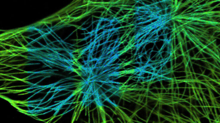
, SPY-Actin (cyan), and SiR-Tubulin (magenta). Instant Computational Clearing (ICC) was applied.")
How to Perform Dynamic Multicolor Time-Lapse Imaging
Live-cell imaging sheds light on diverse cellular events. As many of these events have fast dynamics, the microscope imaging system must be fast enough to record every detail. One major advantage of…
Loading...

Multiplexing through Spectral Separation of 11 Colors
Fluorescence microscopy is a fundamental tool for life science research that has evolved and matured together with the development of multicolor labeling strategies in cells tissues and model…
Loading...
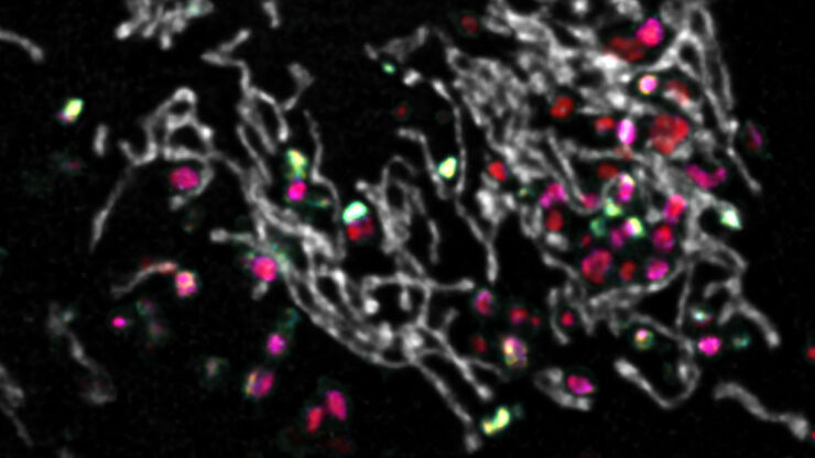
 and CellEvent™ (yellow).")
Following Multiple Events during Staurosporine Apoptosis
In this video on demand, we show how adding additional markers to an apoptosis kit can markedly increase the amount of information a researcher can obtain from the same experiment. The simultaneous…
Loading...

RNA Quality after Different Tissue Sample Preparation
The influence of sample preparation and ultraviolet (UV) laser microdissection (UV LMD) on the quality of RNA from murine-brain tissue cryo-sections is described in this article. To obtain good…
Loading...

New Imaging Tools for Cryo-Light Microscopy
New cryo-light microscopy techniques like LIGHTNING and TauSense fluorescence lifetime-based tools reveal structures for cryo-electron microscopy.
