
Scienze della vita
Scienze della vita
Questo è il posto giusto per ampliare le vostre conoscenze, le capacità di ricerca e le applicazioni pratiche della microscopia in vari campi scientifici. Imparate come ottenere una visualizzazione precisa, l'interpretazione delle immagini e i progressi della ricerca. Troverete informazioni approfondite sulla microscopia avanzata, sulle tecniche di imaging, sulla preparazione dei campioni e sull'analisi delle immagini. Gli argomenti trattati comprendono la biologia cellulare, le neuroscienze e la ricerca sul cancro, con particolare attenzione alle applicazioni e alle innovazioni più avanzate.
Filter articles
Tag
Story Type
Products
Loading...

Organismi Modello nella Ricerca
Un organismo modello è una specie utilizzata dai ricercatori per studiare specifici processi biologici. Hanno caratteristiche genetiche simili a quelle umane e vengono normalmente impiegate in aree di…
Loading...

Virologia
L’oggetto della tua ricerca si concentra su infezioni e malattie virali? Scopri in che modo puoi approfondire l’aspetto virologico utilizzando le soluzioni di imaging e la preparazione del campione di…
Loading...

How Marine Microorganism Analysis can be Improved with High-pressure Freezing
In this application example we showcase the use of EM-Sample preparation with high pressure freezing, freeze substiturion and ultramicrotomy for marine biology focusing on ultrastructural analysis of…
Loading...

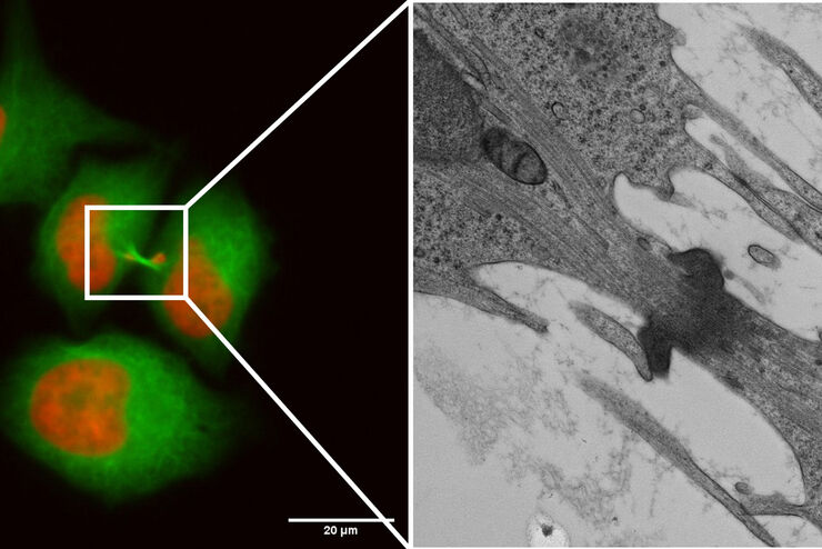
How to Successfully Perform Live-Cell CLEM
The Leica Nano workflow provides a streamlined live-cell CLEM solution for getting insight bout structural changes of cellular components over time. Besides the technical handling described in the…
Loading...

How to Successfully Implement Coral Life
The live-cell CLEM workflow allows you to capture dynamic information related to a relevant biological process as it happens and put these observations into their ultrastructural context. The Leica…
Loading...

How to Improve Live Cell Imaging with Coral Life
For live-cell CLEM applications, light microscopy imaging is a critical step for identifying the right cell in the right state at the right time. In this article, Leica experts share their insights on…
Loading...

How to Keep Your Samples Under Physiological Conditions
The Coral Life workflow combines dynamic data with the best possible sample fixation by high pressure freezing. However, good sample preservation won’t help if your cells are stressed by temperature…
Loading...
Capture life as it happens
With the Leica Nano Workflow, searching for the needle in the haystack is a thing of the past. Take advantage of correlative light and electron microscopy to identify directly the right cell at the…
Loading...
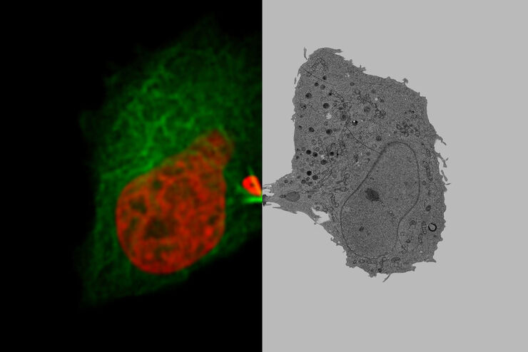
Putting Dynamic Live Cell Data into the Ultrastructural Context
With workflow Coral Life, searching for a needle in the haystack is a thing of the past. Take advantage of correlative light and electron microscopy to identify directly the right cell at the right…
