
is mobile? false
Scienze della vita
Scienze della vita
Questo è il posto giusto per ampliare le vostre conoscenze, le capacità di ricerca e le applicazioni pratiche della microscopia in vari campi scientifici. Imparate come ottenere una visualizzazione precisa, l'interpretazione delle immagini e i progressi della ricerca. Troverete informazioni approfondite sulla microscopia avanzata, sulle tecniche di imaging, sulla preparazione dei campioni e sull'analisi delle immagini. Gli argomenti trattati comprendono la biologia cellulare, le neuroscienze e la ricerca sul cancro, con particolare attenzione alle applicazioni e alle innovazioni più avanzate.
Popolare Show subnavigation
Scienze della vita


Filter articles
Tag
Story Type
Products
Loading...

Live-Cell Fluorescence Lifetime Multiplexing Using Organic Fluorophores
On-demand video: Imaging more subcellular targets by using fluorescence lifetime multiplexing combined with spectrally resolved detection.
Loading...
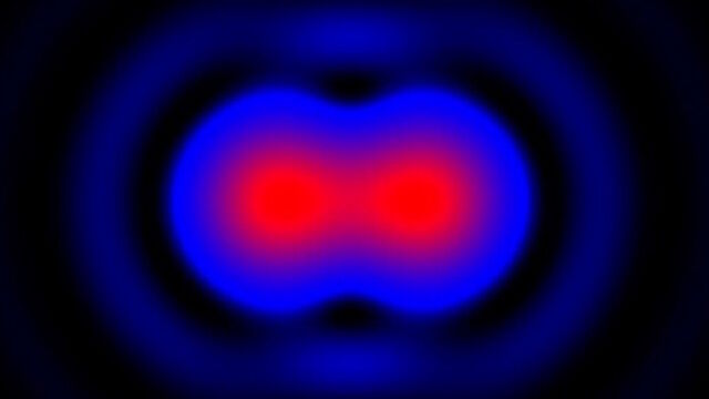
 and acceptor (A) molecule which participate in FRET (Förster resonance energy transfer).")
What is FRET with FLIM (FLIM-FRET)?
This article explains the FLIM-FRET method which combines resonance energy transfer and fluorescence lifetime imaging to study protein-protein interactions.
Loading...
Insights into Vesicle Trafficking
STELLARIS provides integral access to complementary layers of information for dynamic, structural, and mechanistic insights into vesicle trafficking.
Loading...
Visualizing Protein-Protein Interactions by Non-Fitting and Easy FRET-FLIM Approaches
The Webinar with Dr. Sergi Padilla-Parra is about visualizing protein-protein interaction. He gives insight into non-fitting and easy FRET-FLIM approaches.
Loading...

Multiplexing through Spectral Separation of 11 Colors
Fluorescence microscopy is a fundamental tool for life science research that has evolved and matured together with the development of multicolor labeling strategies in cells tissues and model…
Loading...
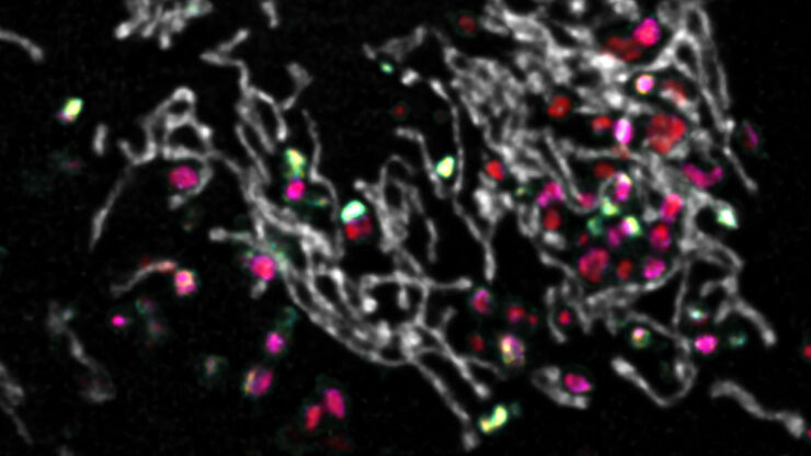
 in 14 x 18 tiles. Lifetime gives an additional contrast that allows to differentiate different structures in histological stainings.")
A Guide to Fluorescence Lifetime Imaging Microscopy (FLIM)
The fluorescence lifetime is a measure of how long a fluorophore remains on average in its excited state before returning to the ground state by emitting a fluorescence photon.
Loading...
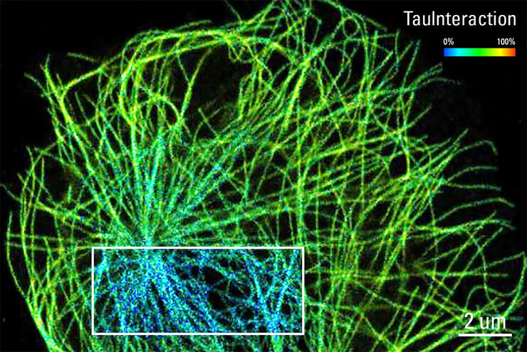
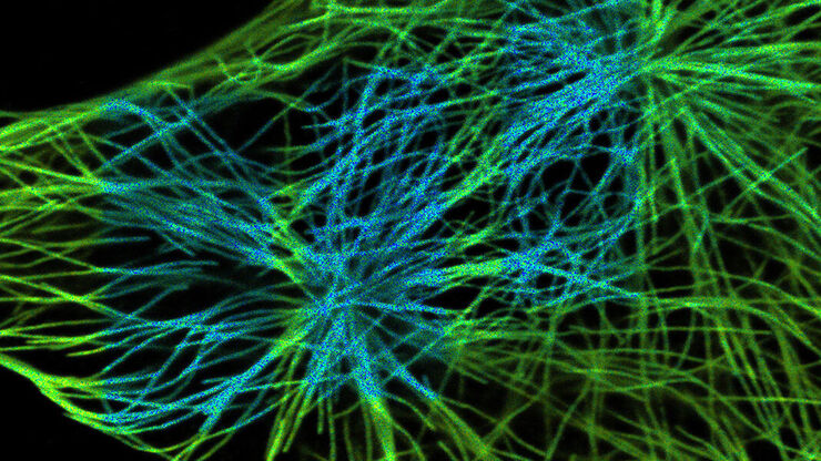
TauInteraction – Studying Molecular Interactions with TauSense
Fluorescence microscopy constitutes one of the pillars in life sciences and is a tool commonly used to unveil cellular structure and function. A key advantage of fluorescence microscopy resides in the…
Loading...

Find Relevant Specimen Details from Overviews
Switch from searching image by image to seeing the full overview of samples quickly and identifying the important specimen details instantly with confocal microscopy. Use that knowledge to set up…
Loading...

How to Prepare your Specimen for Immunofluorescence Microscopy
Immunofluorescence (IF) is a powerful method for visualizing intracellular processes, conditions and structures. IF preparations can be analyzed by various microscopy techniques (e.g. CLSM,…
