
Scienze della vita
Scienze della vita
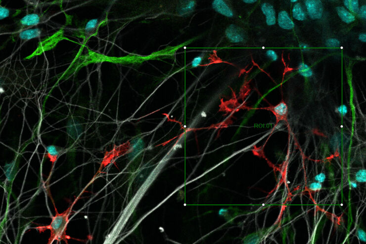
Questo è il posto giusto per ampliare le vostre conoscenze, le capacità di ricerca e le applicazioni pratiche della microscopia in vari campi scientifici. Imparate come ottenere una visualizzazione precisa, l'interpretazione delle immagini e i progressi della ricerca. Troverete informazioni approfondite sulla microscopia avanzata, sulle tecniche di imaging, sulla preparazione dei campioni e sull'analisi delle immagini. Gli argomenti trattati comprendono la biologia cellulare, le neuroscienze e la ricerca sul cancro, con particolare attenzione alle applicazioni e alle innovazioni più avanzate.
Filter articles
Tag
Story Type
Products
Loading...
Explore Innovative Techniques to Separate Fluorophores with Overlapping Spectra
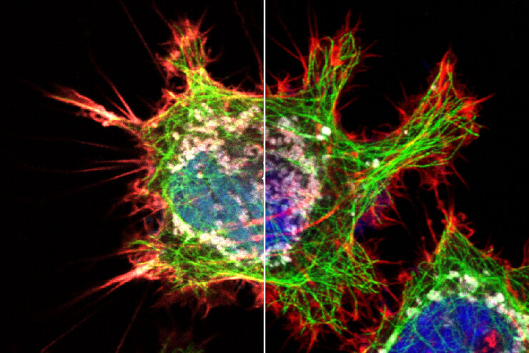
In this article we explore several strategies you can take to improve the separation of fluorophores and increase the number of fluorescent probes you can distinguish in your sample.
Loading...

STELLARIS White Light Lasers
When it comes to choosing fluorescent probes for your multi-color experiments, you shouldn’t have to compromise. Now you can advance beyond conventional excitation sources that limit your fluorophore…
Loading...
TauSense Technology Imaging Tools
Leica Microsystems’ TauSense technology is a set of imaging modes based on fluorescence lifetime. Found at the core of the STELLARIS confocal platform, it will revolutionize your imaging experiments.…
Loading...

The Power HyD Detector Family
Powerful photon counting detectors on the STELLARIS confocal platform provide improved photon counting, ultra-sensitive imaging and more color options in the NIR spectrum.
Loading...
 from the desired fluorescent signal from the cell membrane (green).")
Learn how to Remove Autofluorescence from your Confocal Images
Autofluorescence can significantly reduce what you can see in a confocal experiment. This article explores causes of autofluorescence as well as different ways to remove it, from simple media fixes to…
Loading...

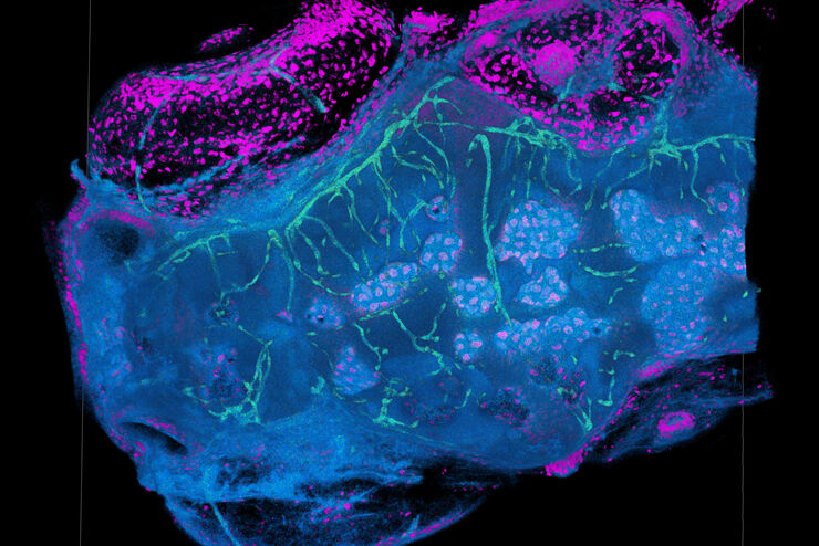
Zebrafish Brain - Whole Organ Imaging at High Resolution
Structural information is key when one seeks to understand complex biological systems, and one of the most complex biological structures is the vertebrate central nervous system. To image a complete…
Loading...
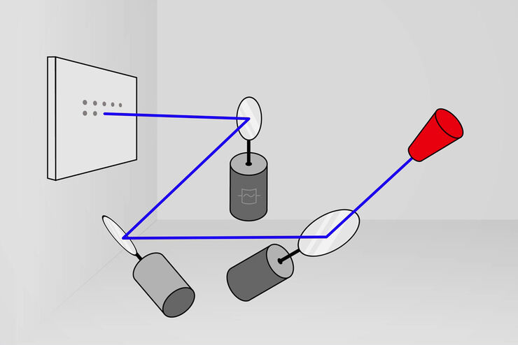
What is a Resonant Scanner?
A resonant scanner is a type of galvanometric mirror scanner that allows fast image acquisition with single-point scanning microscopes (true confocal and multiphoton laser scanning). High acquisition…
Loading...
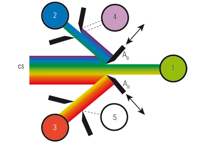
What is a Spectral Detector (SP Detector)?
The SP detector from Leica Microsystems denotes a compound detection unit for point scanning microscopes, in particular confocal microscopes. The SP detector splits light into up to 5 spectral bands.…
Loading...
Resolved Field Number (RFN)
The field number (FN) for optical microscopes indicates the field of view (FOV). It corresponds to the area in the intermediate image that is observable through the eyepieces. Although, we cannot…
