
Science Lab
Science Lab
Benvenuti nel portale delle conoscenze di Leica Microsystems. Troverete materiale didattico e di ricerca scientifica sul tema della microscopia. Il portale supporta i principianti, i professionisti esperti e gli scienziati nel loro lavoro quotidiano e negli esperimenti. Esplorate i tutorial interattivi e le note applicative, scoprite le basi della microscopia e le tecnologie di punta. Entrate a far parte della comunità di Science Lab e condividete la vostra esperienza.
Filter articles
Tag
Story Type
Products
Loading...

Optimizing THUNDER Platform for High-Content Slide Scanning
With rising demand for full-tissue imaging and the need for FL signal quantitation in diverse biological specimens, the limits on HC imaging technology are tested, while user trainability and…
Loading...
Visualizing Protein Degradation and Aggregation in the Living Cell
Our guest speaker, Prof Dr Eric Reits, presents his work on neurodegenerative disorders. Reits’ group are experts on the subject of Huntington’s disease and work towards identifying leads for…
Loading...
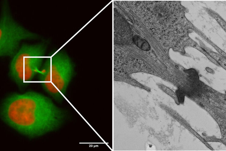
Capture life as it happens
With the Leica Nano Workflow, searching for the needle in the haystack is a thing of the past. Take advantage of correlative light and electron microscopy to identify directly the right cell at the…
Loading...
Life Beyond the Pixels: Deep Learning Methods for Single Cell Analysis
Our guest speaker Prof Dr Peter Horvath presents his work on single cell-based large-scale microscopy experiments. This novel targeting approach includes the use of machine learning models and…
Loading...

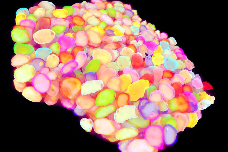
Tissue Image Gallery
Visual analysis of animal and human tissues is critical to understand complex diseases such as cancer or neurodegeneration. From basic immunohistochemistry to intravital imaging, confocal microscopy…
Loading...
, TL DIC")
DIC
Un microscopio DIC è basato su un microscopio composto dotato di un filtro di polarizzazione e di un prisma di Wollaston posizionati rispettivamente tra la sorgente luminosa e la lente del…
Loading...

Dissecting Proteomic Heterogeneity of the Tumor Microenvironment
This lecture will highlight cutting edge applications in applying laser microdissection and microscaled quantitative proteomics and phosphoproteomics to uncover exquisite intra- and inter-tumor…
Loading...

Microscopi per campo scuro
Il metodo di contrasto in campo oscuro sfrutta la diffrazione o la dispersione della luce da strutture di un campione biologico o da caratteristiche non uniformi nella struttura di un materiale.
Loading...
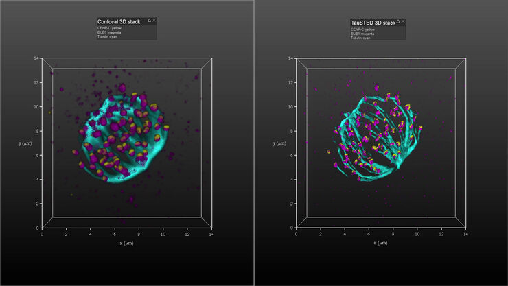
Kinetochore Assembly during Mitosis with TauSTED on 3D
Three-dimensional organization of the mitotic spindle together with the distribution of CENP-C and BUB1 based on TauSTED with multiple STED lines (592, 660 and 775 nm) can provide insights…
