不明点・ご質問は、アプリケーションのスペシャリストへお気軽にご相談ください。
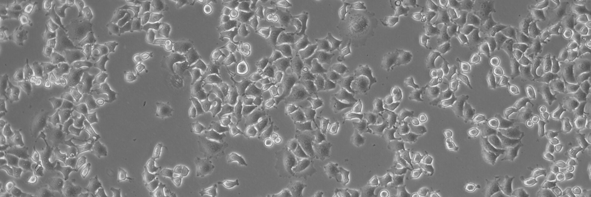
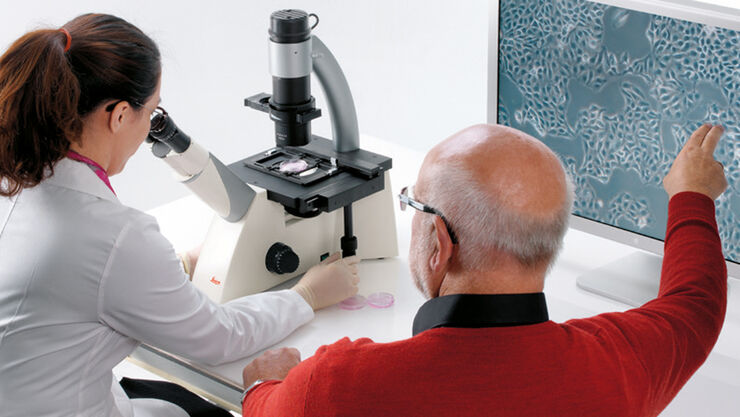
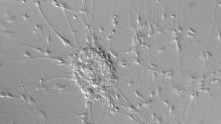
動物細胞は本質的にコントラストが非常に低いために、培養細胞用顕微鏡は位相差コントラストなどの観察法をサポートできる必要があります。また、培養に使用している容器はプラスチック製であることが多く、その場合は DIC(微分干渉コントラスト)が使えません。一方、DIC に代わる優れた手段として IMC モジュラーコントラストが用いられます。これは、プラスチック容器に対しても観察が可能であり、かつ専用の対物レンズやプリズムを用意する必要がありません。培養細胞用顕微鏡は、さらに作業効率をアップさせるために、取り扱いが容易である必要があります。
ライカの培養細胞用顕微鏡は使いやすいうえに、個々の目的に合わせてユーザーが選択した観察法に柔軟に対応します。
チュートリアル
Phase Contrast
Phase contrast is an optical contrast technique for making unstained phase objects (e.g. flat cells) visible under the optical microscope. Cells that appear inconspicuous and transparent in brightfield can be viewed in high contrast and rich detail using a phase contrast microscope.
Differential Interference Contrast
Differential interference contrast (DIC) microscopy is a good alternative to brightfield microscopy for gaining proper images of unstained specimens that often only provide a weak image in brightfield.
Integrated Modulation Contrast
Hoffman modulation contrast has established itself as a standard for the observation of unstained, low-contrast biological specimens. Its innovative technical implementation permits significantly simpler handling and greater flexibility in deployment.
{{ question.questionText }}
答えを選んでください!
ベストマッチ
{{ resultProduct.header }}
{{ resultProduct.subheader }}
{{ resultProduct.description }}
{{ resultProduct.features }}
資料請求
細胞培養用製品
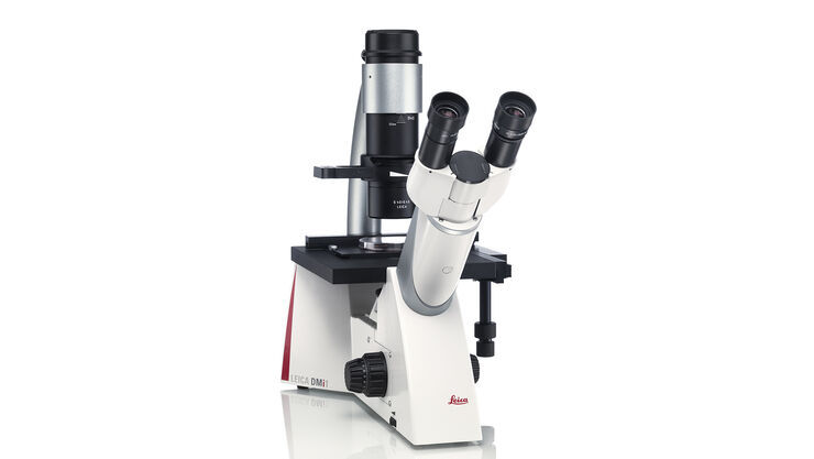
細胞培養・組織培養用ルーチン倒立顕微鏡 ライカ Leica DMi1
ライカ DMi1 倒立顕微鏡はルーチン作業をサポートします。直観的に操作でき、楽に扱えるので、作業に集中することができます。各種アクセサリも用意されており、作業にとって重要なアイテムを容易に追加することができます。
研究用倒立顕微鏡 Leica DM IL LED
Leica DM IL LEDは、お客様のニーズに合ったコントラスト法の包括的なセットを備えています。 高品質の位相差コントラスト、優れた変調コントラスト、高輝度の蛍光観察にすぐに利用できます。 堅牢な安定性、広い作業スペース、大型の培養フラスコ観察に対応した長い作動距離、熱線フリーの安定した照明により、顕微鏡上での作業が簡単かつ快適になります。
| 明視野 | 位相差コントラスト法(PH) | 微分干渉コントラスト法(DIC) | 積分変調コントラスト法(IMC) | 蛍光 | 倍率 | 作動距離 | カメラ | |
Leica DM IL LED | + | + | - | + | + | PH: 5x to 63x IMC: 10x, 20x, 32x, 40x | 40 mm, 80 mm | + (自由選択) |
Leica DMi1 | + | + | - | - | - | 10x, 20x, 40x | 40 mm, 50 mm, 80 mm | + (統合) |
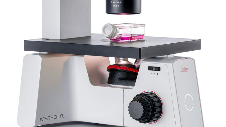
Mateo TL | + | + | - | - | - | 4x, 10x, 20x, 40x | 50 mm | +(統合) |
Mateo FL | + | + | - | - | + | 2.5x, 4x, 5x, 10x, 20x, 40x, 63x | 50 mm | + デュアルカメラ (統合) |
培養細胞観察専用の顕微鏡。
顕微鏡 – より高度な要求に応える
どのようなツールが必要でしょうか?
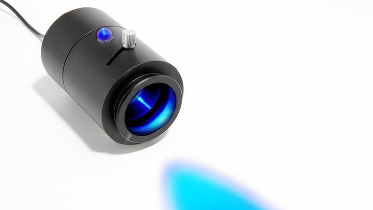
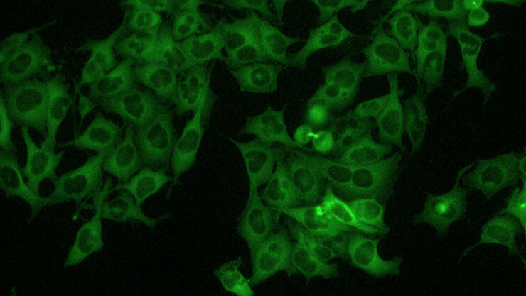
細胞生物学研究の現場では、細胞に蛍光マーカーを導入し、その後の挙動を研究用顕微鏡で観察する手法が広く用いられます。トラッキングの対象が蛍光タンパク質である場合、培養細胞用顕微鏡でトランスフェクション効率などを確認するために蛍光オプションが利用可能であることも必要です。
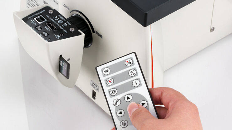
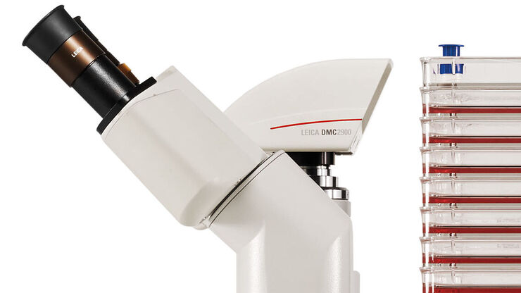
レポート作成とその作業の標準化として、顕微鏡にデジタルカメラを装備し、取得画像とそれにまつわるデータを取りまとめる機能も必要です。
スペースの問題と常に向き合う研究室では、培養細胞用顕微鏡はフードの中に収まることが期待されます。近年では、インキュベーター内に入れて使用できるほど、小型かつ堅牢な顕微鏡が求められる傾向にあります。
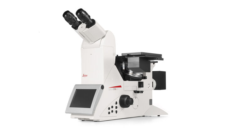
シャーレ内で単一細胞の変化を精密に観察したい、複数の分析手法でスクリーニングしたい、単一分子の解像度を取得したい、または複雑なプロセスの中での細胞のふるまいを紐解きたい時にも、DMi8 S システムなら、より鮮明に、より早く、核心にたどりつくことが出来ます。
チュートリアル
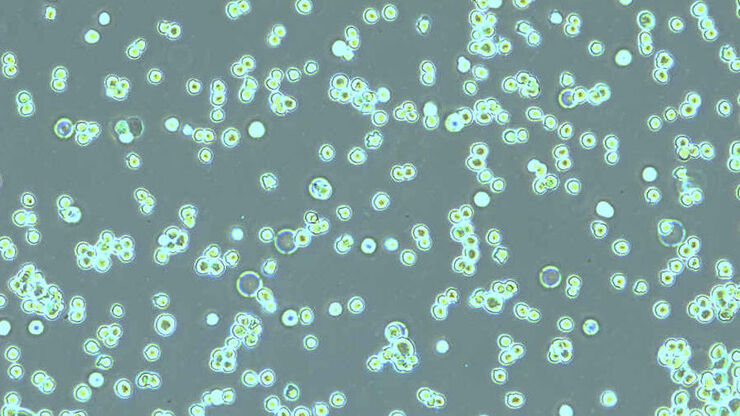
How to do a Proper Cell Culture Quick Check
Many fields of biomedical research, like cancer research, drug development and tissue engineering, require the use of living cells to perform a variety of assays. Mammalian cell cultures are an essential tool in biology because they allow rapid growth and proliferation of different cell types for experimental analysis.
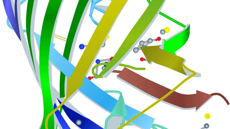
Fluorescent Proteins
The prospects of fluorescence microscopy changed dramatically with the discovery of fluorescent proteins in the 1950s. The starting point was the detection of the jellyfish Aequorea victoria green fluorescent protein (GFP) by Osamo Shimomura. Hundreds of GFP mutants later, the range of fluorescent proteins reaches from the blue to the red spectrum.
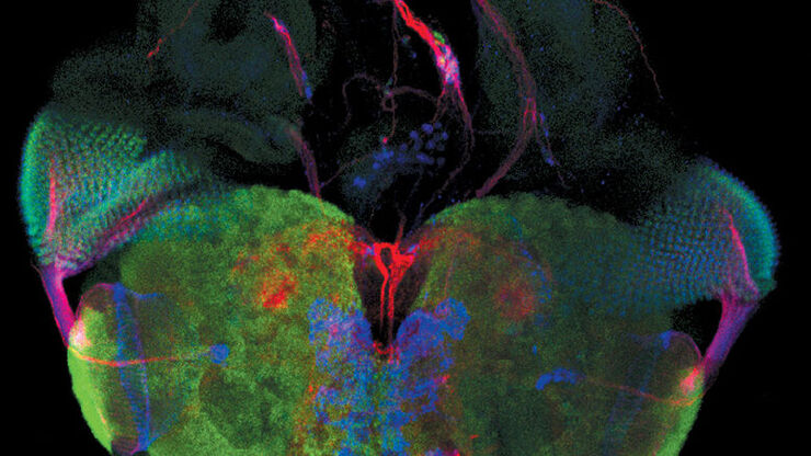
An Introduction to Fluorescence
Fluorescence is an effect which was first described by George Gabriel Stokes in 1852. He observed that fluorite begins to glow after being illuminated with ultraviolet light. Fluorescence is a form of photoluminescence which describes the emission of photons by a material after being illuminated with light. The emitted light is of longer wavelength than the exciting light. This effect is called the Stokes shift.
