K3C & K3M
デジタルカメラ
製品紹介
Home
Leica Microsystems
K3C & K3M 顕微鏡用カメラ シリーズ
ライフサイエンスおよび産業用 画像解析ソリューション
最新の記事を読む
がん研究
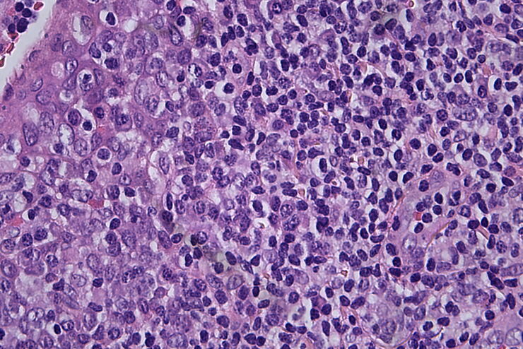
がんは、成長調節における欠損細胞によって引き起こされる複雑な異質性疾患です。 細胞または細胞群内の遺伝的および後成的変化が通常の機能を妨げ、自律的、非制御の細胞成長と増殖を引き起こします。
Technical Terms for Digital Microscope Cameras and Image Analysis
Learn more about the basic principles behind digital microscope camera technologies, how digital cameras work, and take advantage of a reference list of technical terms from this article.
Understanding Clearly the Magnification of Microscopy
To help users better understand the magnification of microscopy and how to determine the useful range of magnification values for digital microscopes, this article provides helpful guidelines.
Life Science Research: Which Microscope Camera is Right for You?
Deciding which microscope camera best fits your experimental needs can be daunting. This guide presents the key factors to consider when selecting the right camera for your life science research.
Factors to Consider when Selecting Clinical Microscopes
What matters if you would like to purchase a clinical microscope? Learn how to arrive at the best buying decision from our Science Lab Article.
Clinical Microscopy: Considerations on Camera Selection
The need for images in pathology laboratories has significantly increased over the past few years, be it in histopathology, cytology, hematology, clinical microbiology, or other applications. They…
The Time to Diagnosis is Crucial in Clinical Pathology
Abnormalities in tissues and fluids - that’s what pathologists are looking for when they examine specimens under the microscope. What they see and deduce from their findings is highly influential, as…
応用分野
ライフサイエンス
ライカマイクロシステムズのライフサイエンスリサーチ部門は、革新的技術と理化学分野の深い専門知を要する業界において、微細構造の可視化、測定、分析のためのイメージングソリューションを提供します。
インダストリーマーケット別
稼働時間を最大化し、生産性向上により、お客様の収益に貢献します。ライカの顕微鏡(マイクロスコープ)ソリューションは、微細な異物や残渣なども見逃しなく、迅速かつ信頼性の高い分析、文書化、結果報告を実現します。ライカマイクロシステムズは、幅広いソリューションとエキスパートによるサポートを提供し、さまざまなアプリケーションのニーズにお応えします。
