Leica DM2500 & DM2500 LED
正立顕微鏡
光学顕微鏡
製品紹介
Home
Leica Microsystems
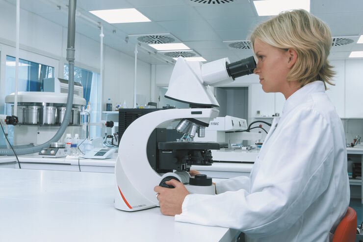
Leica DM2500 & DM2500 LED 強力な照明を備えた、人間工学に基づく先進の顕微鏡システム
最新の記事を読む
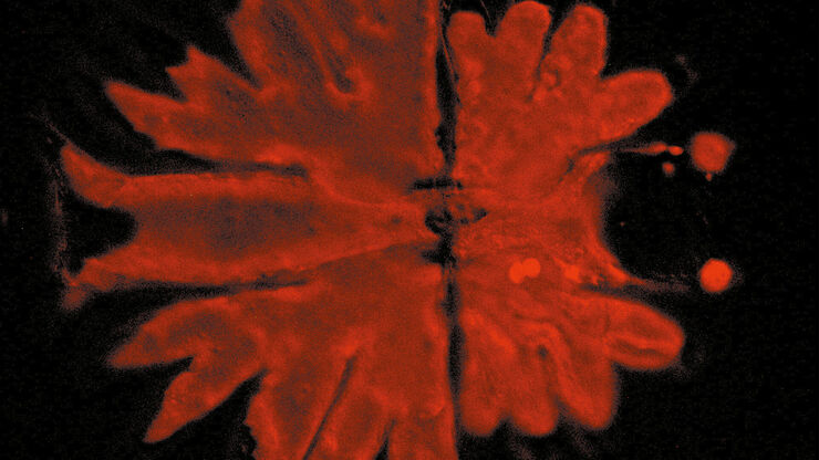
位相コントラスト
位相差顕微鏡では、染色することなく、多くの種類の生物試料の構造をより優れたコントラストで観察できます。
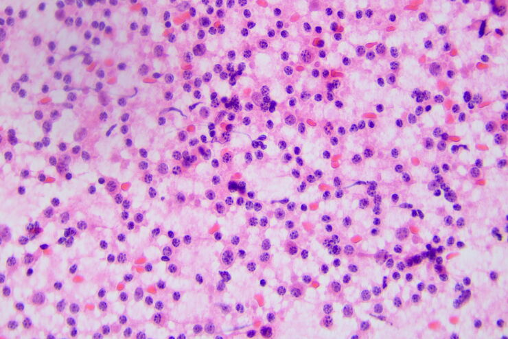
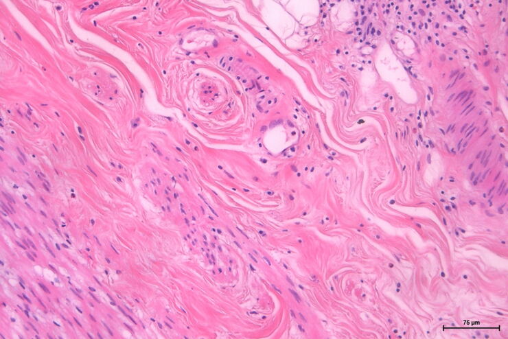
H&E Staining in Microscopy
If we consider the role of microscopy in pathologists’ daily routines, we often think of the diagnosis. While microscopes indeed play a crucial role at this stage of the pathology lab workflow, they…
How to Benefit from Digital Cytopathology
If you have thought of digital cytopathology as characterized by the digitization of glass slides, this webinar with Dr. Alessandro Caputo from the University Hospital of Salerno, Italy will broaden…
Factors to Consider when Selecting Clinical Microscopes
What matters if you would like to purchase a clinical microscope? Learn how to arrive at the best buying decision from our Science Lab Article.
The Time to Diagnosis is Crucial in Clinical Pathology
Abnormalities in tissues and fluids - that’s what pathologists are looking for when they examine specimens under the microscope. What they see and deduce from their findings is highly influential, as…
微分干渉(DIC)顕微鏡
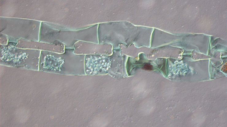
微分干渉(DIC)顕微鏡は、光源と集光レンズの間に偏光フィルターとウォラストンプリズムを配置した広視野顕微鏡で、対物レンズとカメラセンサーや接眼レンズの間にも偏光フィルターとウォラストンプリズムを配置しています。
暗視野顕微鏡
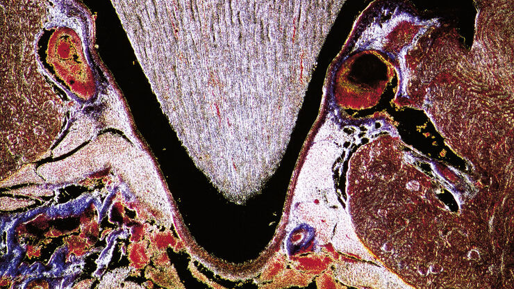
暗視野コントラスト法は、生体試料の構造や物質試料の不均一な特徴から生じる光の回折や散乱を利用する方法です。
Koehler Illumination: A Brief History and a Practical Set Up in Five Easy Steps
In this article, we will look at the history of the technique of Koehler Illumination in addition to how to adjust the components in five easy steps.
Forensic Detection of Sperm from Sexual Assault Evidence
The impact of modern scientific methods on the analysis of crime scene evidence has dramatically changed many forensic sub-specialties. Arguably one of the most dramatic examples is the impact of…
応用分野
病理研究
ライカの病理顕微鏡ソリューションをご紹介します。
病理向けソリューション
病理学の試料分析では、長時間にわたり顕微鏡を使用する必要があります。 顕微鏡の利用者にストレスや身体的な負担をかけ、効率性の低下につながる恐れがあります。 ライカの顕微鏡は快適な姿勢を保ち、負担を軽減するソリューションを提供します 病理診断の効率性向上に貢献します
解剖病理学
ライカマイクロシステムズの病理用顕微鏡が、効率的で正確な医療診断をいかにサポートするかをご紹介します。
