Viventis Deep
ライトシート顕微鏡
製品紹介
Home
Leica Microsystems
Viventis Deep デュアルビューライトシート顕微鏡
生命の全容を明らかにする
最新の記事を読む
How to Study Gene Regulatory Networks in Embryonic Development
Join Dr. Andrea Boni by attending this on-demand webinar to explore how light-sheet microscopy revolutionizes developmental biology. This advanced imaging technique allows for high-speed, volumetric…
Dual-View LightSheet Microscope for Large Multicellular Systems
Visualizing the dynamics of complex multicellular systems is a fundamental goal in biology. To address the challenges of live imaging over large spatiotemporal scales, Franziska Moos et. al. present…
Download The Guide to Live Cell Imaging
In life science research, live cell imaging is an indispensable tool to visualize cells in a state as in vivo as possible. This E-book reviews a wide range of important considerations to take to…
研究におけるモデル生物
モデル生物とは、特定の生物学的プロセスを研究するために研究者が使用する生物種です。 モデル生物は、人間と似た遺伝的特徴を持ち、遺伝子学、発生生物学、神経科学などの研究分野で一般的に使用されています。 通常、モデル生物は実験環境での維持や繁殖が容易であること、生殖サイクルが短いこと、または、特定の形質や病気を研究するために突然変異体を生成する能力を持つことで選ばれます。
応用分野
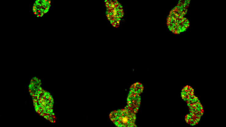
オルガノイドと3D細胞培養
ライフサイエンス研究で近年最も目覚ましい発展の一つは、オルガノイド、スフェロイド、生体機能チップモデルなどの3D細胞培養システムの開発です。 3D細胞培養は、細胞が成長し、全3次元で周囲と相互作用できる人工環境です。 これらの条件は、生体内条件に似ています。


