THUNDER Imaging Systems

THUNDER Imaging Systems

THUNDER Imaging Systems

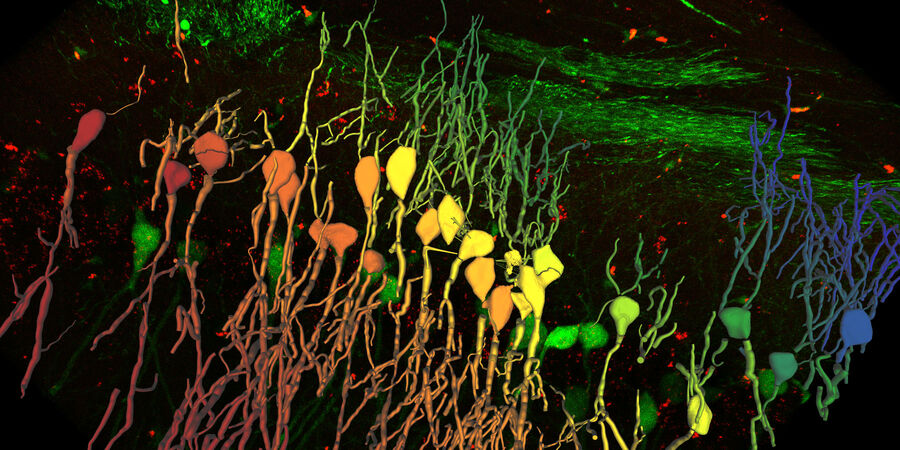
このシステムは、サンプルの深部も非侵襲でボケのない鮮明な画像をリアルタイムに提供し、生体の極めて重要な現象に答える助けとなります。カメラベースの蛍光顕微鏡による従来の作業と同様の簡便さで 3D サンプルの鮮明なイメージングを実現します。Computational Clearing 機能を持つ THUNDER システムは、厚みのある立体的なサンプルの高速、高品質イメージングのための新しい装置です。

THUNDER モデル生物

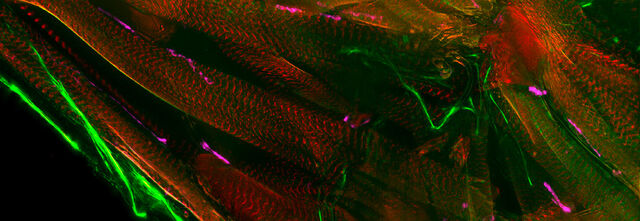
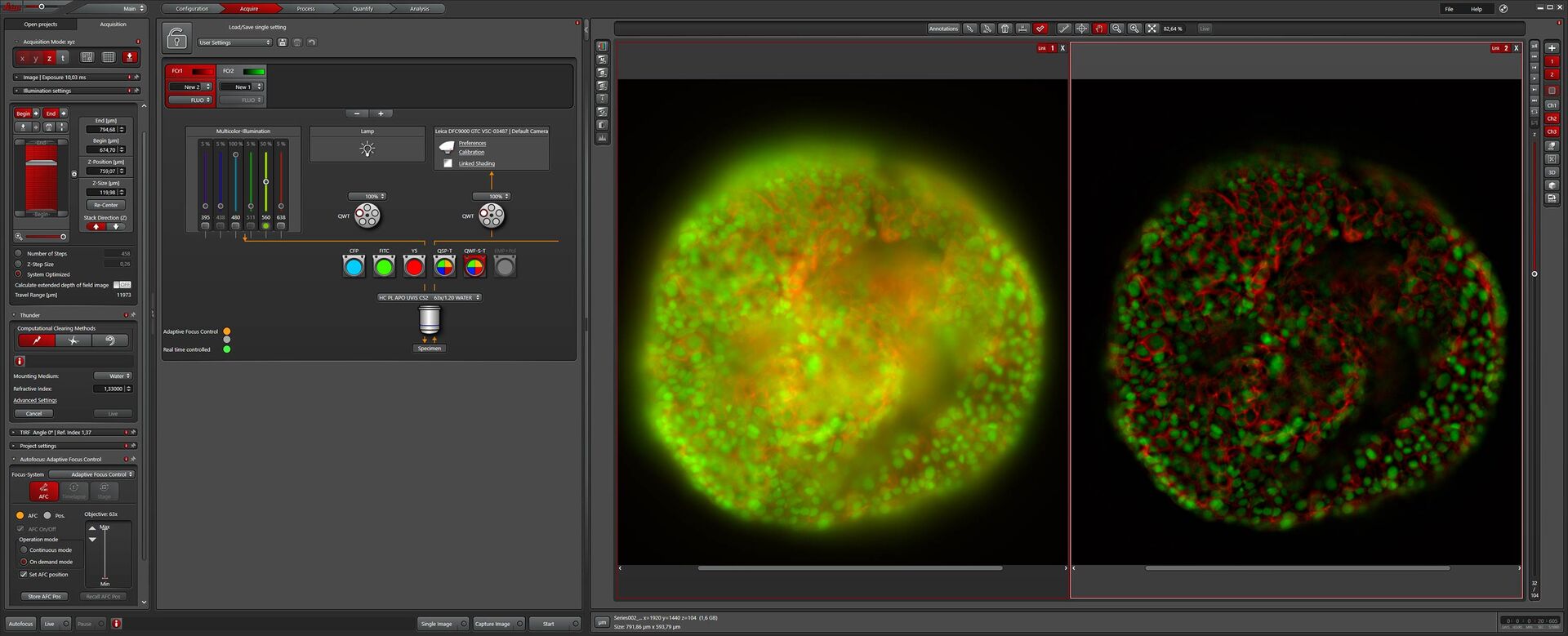
THUNDER モデル生物を用いると、発生生物学・分子生物学研究を目的に、素早く簡単に 3D 観察することができます。
THUNDER 組織標本

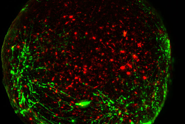
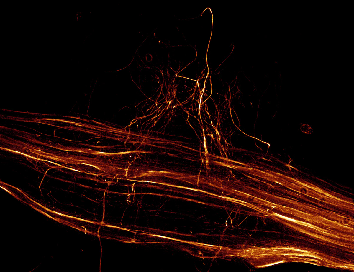
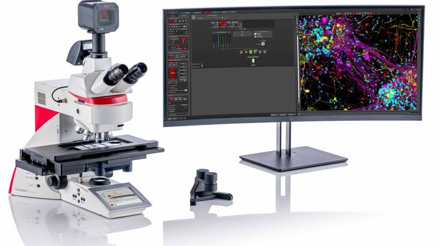
THUNDER 組織標本では、神経科学や組織学研究でよく用いられる組織切片の三次元の蛍光像をリアルタイムに取得することができます。
THUNDER 3D 生細胞および 3D 培養細胞

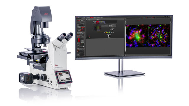
THUNDER は幹細胞、スフェロイド、オルガノイドなど、高度な 3 次元培養アッセイのためのソリューションです。

Read our latest articles about THUNDER Imaging Systems

The knowledge portal of Leica Microsystems offers scientific research and teaching material on the subjects of microscopy. The content is designed to support beginners, experienced practitioners and scientists alike in their everyday work and experiments.
その他の記事

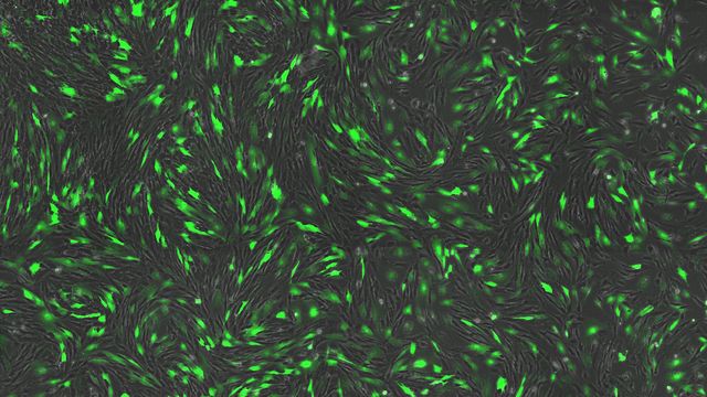
Designing the Future with Novel and Scalable Stem Cell Culture
Visionary biotech start-up Uncommon Bio is tackling one of the world’s biggest health challenges: food sustainability. In this webinar, Stem Cell Scientist Samuel East shows how they make stem cell culture media for cellular agriculture safe and economically viable. See how they achieved a 1000x reduction in media costs and developed animal free, food safe iPSC culture media.

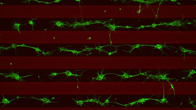
Revealing Neuronal Migration’s Molecular Secrets

Different approaches can be used to investigate neuronal migration to their niche in the developing brain. In this webinar, experts from The University of Oxford present the microscopy tools and assays they use to elucidate the molecular mechanisms of neuronal migration to functional layers of the cortex during neurodevelopment. Understanding these processes will lead to a better understanding of healthy brain development and potentially better treatments for neurodevelopmental disorders.

How Efficient is your 3D Organoid Imaging and Analysis Workflow?

Organoid models have transformed life science research but optimizing image analysis protocols remains a key challenge. This webinar explores a streamlined workflow for organoid research, starting with real-time 3D cell culture checks. This is followed by high-speed, high-resolution 3D imaging to generate crisp images and cleaner data for accurate AI-based segmentation and quantification of parameters such as growth rate, cell migration and 3D cellular interactions – enabling deeper insights.

How to Get Deeper Insights into your Organoid and Spheroid Models

In this eBook, learn about key considerations for imaging 3D cultures, such as organoids and spheroids, and discover microscopy solutions to shed new insights into dynamic processes in 3D real-time

Imaging Organoid Models to Investigate Brain Health

Imaging human brain organoid models to study the phenotypes of specialized brain cells called microglia, and the potential applications of these organoid models in health and disease.

機械受容性経路とシナプス経路の研究に顕微鏡がいかに役立つか

このポッドキャストでは、Tobi Langenhan教授は、顕微鏡を使ってシナプスのタンパク質集合体を調べるなど、接着型GPCRの機械受容特性の研究を通して、タンパク質のダイナミクスとその空間的相互作用に精通されています。 Abdullah Ahmed,博士との対談では、Tobi博士と彼のグループが答えようとしている主要な質問と、彼らが直面しているいくつかの課題について話します。彼はまた、THUNDER Imagerテクノロジーを使用して無傷のショウジョウバエの幼虫を画像化したときに得た驚きを共有しています。

What are the Challenges in Neuroscience Microscopy?

eBook outlining the visualization of the nervous system using different types of microscopy techniques and methods to address questions in neuroscience.

The Role of Iron Metabolism in Cancer Progression

Iron metabolism plays a role in cancer development and progression, and modulates the immune response. Understanding how iron influences cancer and the immune system can aid the development of new therapeutic approaches.

Going Beyond Deconvolution

Widefield fluorescence microscopy is often used to visualize structures in life science specimens and obtain useful information. With the use of fluorescent proteins or dyes, discrete specimen components are marked in a highly specific manner. To fully understand a structure, visualizing it in 3 dimensions can be necessary, but certain challenges are faced when doing so with microscopy.

Find Relevant Specimen Details from Overviews

Switch from searching image by image to seeing the full overview of samples quickly and identifying the important specimen details instantly with confocal microscopy. Use that knowledge to set up high-resolution image acquisition automatically using templates for slides, dishes, and multi-well plates. Like a GPS for the cells of a specimen, users always have a clear path to high-quality data with the LAS X Navigator software, a powerful navigation tool for the STELLARIS platform. LAS X Navigator enables confocal and fluorescence lifetime experiments to be combined with a stage application and makes all the TauSense tools available for use.

Accurately Analyze Fluorescent Widefield Images

The specificity of fluorescence microscopy allows researchers to accurately observe and analyze biological processes and structures quickly and easily, even when using thick or large samples. However, out-of-focus fluorescence increases background, reduces contrast, and makes accurate image segmentation more challenging. A powerful solution to these issues is the use of THUNDER to remove image blur in combination with Aivia which automates widefield image analysis using AI to improve speed and accuracy. Here, we explore this synergetic approach in more detail.

High-resolution 3D Imaging to Investigate Tissue Ageing

Award-winning researcher Dr. Anjali Kusumbe demonstrates age-related changes in vascular microenvironments through single-cell resolution 3D imaging of young and aged organs.

How to Improve Live Cell Imaging with Coral Life

For live-cell CLEM applications, light microscopy imaging is a critical step for identifying the right cell in the right state at the right time. In this article, Leica experts share their insights on the advantages of working with a widefield system and which obstacles need to be overcome when using sapphire as a cell-culture substrate.

Optimizing THUNDER Platform for High-Content Slide Scanning
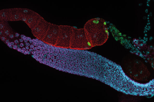
With rising demand for full-tissue imaging and the need for FL signal quantitation in diverse biological specimens, the limits on HC imaging technology are tested, while user trainability and ease-of-use at core facilities becomes a matter of cost and efficiency. Here we demonstrate a feasible workflow developed for the THUNDER platform at our facilities, to support diverse research environments with needs ranging from KO-mouse tissue analysis to human cancer.

Physiology Image Gallery

Physiology is about the processes and functions within a living organism. Research in physiology focuses on the activities and functions of an organism’s organs, tissues, or cells, including the physical and chemical phenomena involved. Here we show you images related to physiology using different samples as examples.

Neuroscience Images

Neuroscience commonly uses microscopy to study the nervous system’s function and understand neurodegenerative diseases.

THUNDER システム

生物学的に重要な 3D モデルを容易に扱えるイメージングシステム:THUNDER システムが登場しました。

このシステムは、サンプルの深部も非侵襲でボケのない鮮明な画像をリアルタイムに提供し、生体の極めて重要な現象に答える助けとなります。カメラベースの蛍光顕微鏡による従来の作業と同様の簡便さで 3D サンプルの鮮明なイメージングを実現します。Computational Clearing 機能を持つ THUNDER システムは、厚みのある立体的なサンプルの高速、高品質イメージングのための新しい装置です。

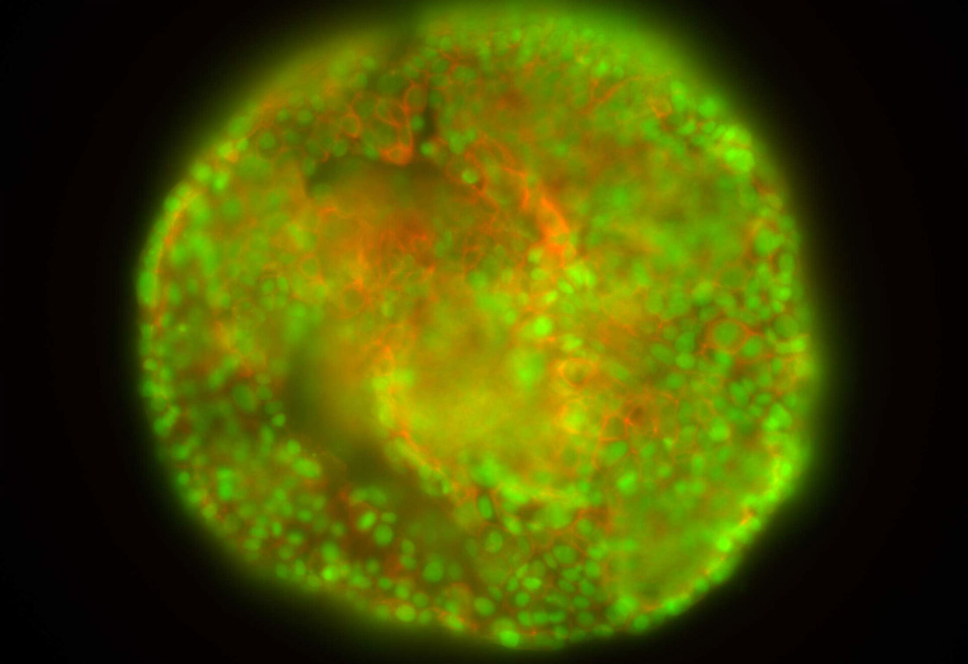
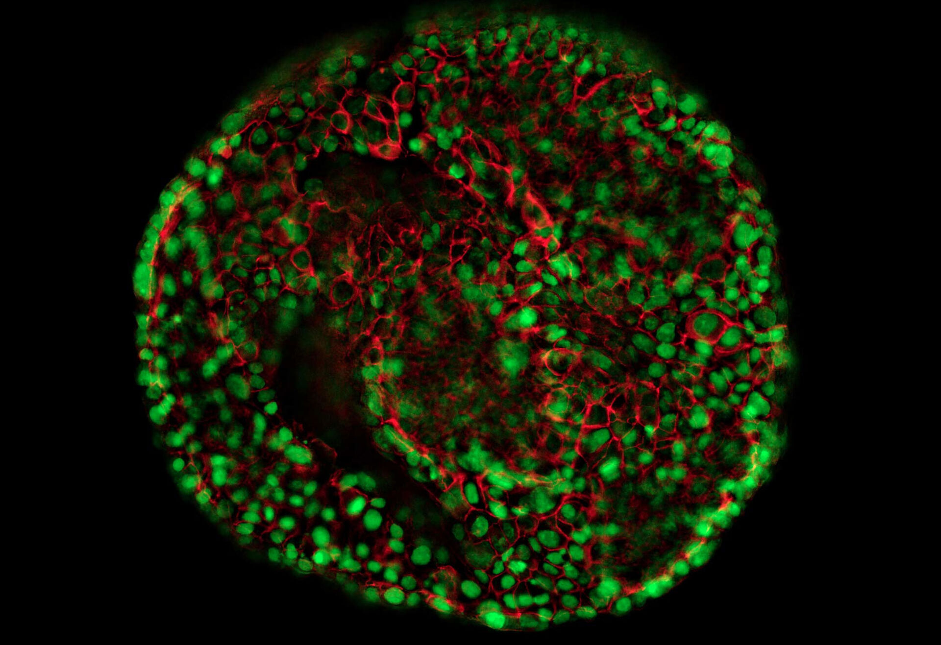
従来の蛍光画像THUNDER画像

HeLa 細胞(標識;アクチン_Alexa Fluor 568 ファロイジン、核_YOYO 1 iodide)

3D バイオロジーをリアルタイムに解き明かす *

モデル生物、組織切片、オルガノイドなどの 3 次元細胞構造の研究方法が THUNDER により大きく変わります。シングルセル、組織、生体全体、腫瘍のスフェロイドのいずれであっても、THUNDER ならば 3D バイオロジーのリアルタイムでの解明が可能です。

THUNDER システムの特長

  • 各アプリケーションのベンチマークとなる最高品質の結果を取得
  • Computational Clearing により、サンプルの深部であっても精細に観察
  • 扱いやすいソフトウェア、速度、感度


*ISO/IEC 2382:2015 準拠

image analysis software solutions

画像を分析し、隠れた洞察を得る

ライカマイクロシステムズが提供する先進の画像解析ソフトウェアソリューションAiviaとLAS Xは、顕微鏡を使った研究を次のレベルへ引き上げるための力を与えてくれます。THUNDER とライカの AI を活用したソフトウェアAivia を組み合わせることで、機械学習や深層学習の幅広いアルゴリズムにより、顕微鏡のイメージングデータから有意義な洞察を得ることができます。

ライカマイクロシステムズの画像解析ソリューションの詳細

今後開催予定のカンファレンス、学会、展示会、オンラインセミナー、ワークショップの情報です。みなさまとお会い出来る機会を楽しみにしております。

すべてのイベント

03
06 Apr 2025

APAO-AIOC 2025

India Congress
05
06 Apr 2025

2025 Singapore ENT-HNS Congress

Singapore Congress

THUNDER の技術

THUNDER は、最新の Computational Clearing 法を用いて高解像度、高コントラストの画像を計算処理で提供するデジタルオプティクス技術です。Computational Clearing は、厚いサンプルでしばしば見られるフォーカスアウトしたボケ情報を除去します。これにより、z スタックでも深部の一平面画像でも優れた結果が得られます。

ライカの THUNDER テクノロジーは、ボケのない画像をリアルタイムに取得するため、関連する光学的パラメータをすべて考慮します。

テクノロジーノート

まだ疑問な点がありますか?THUNDERテクノロジーに関するより詳細な解説は、テクノロジーノートをご覧ください

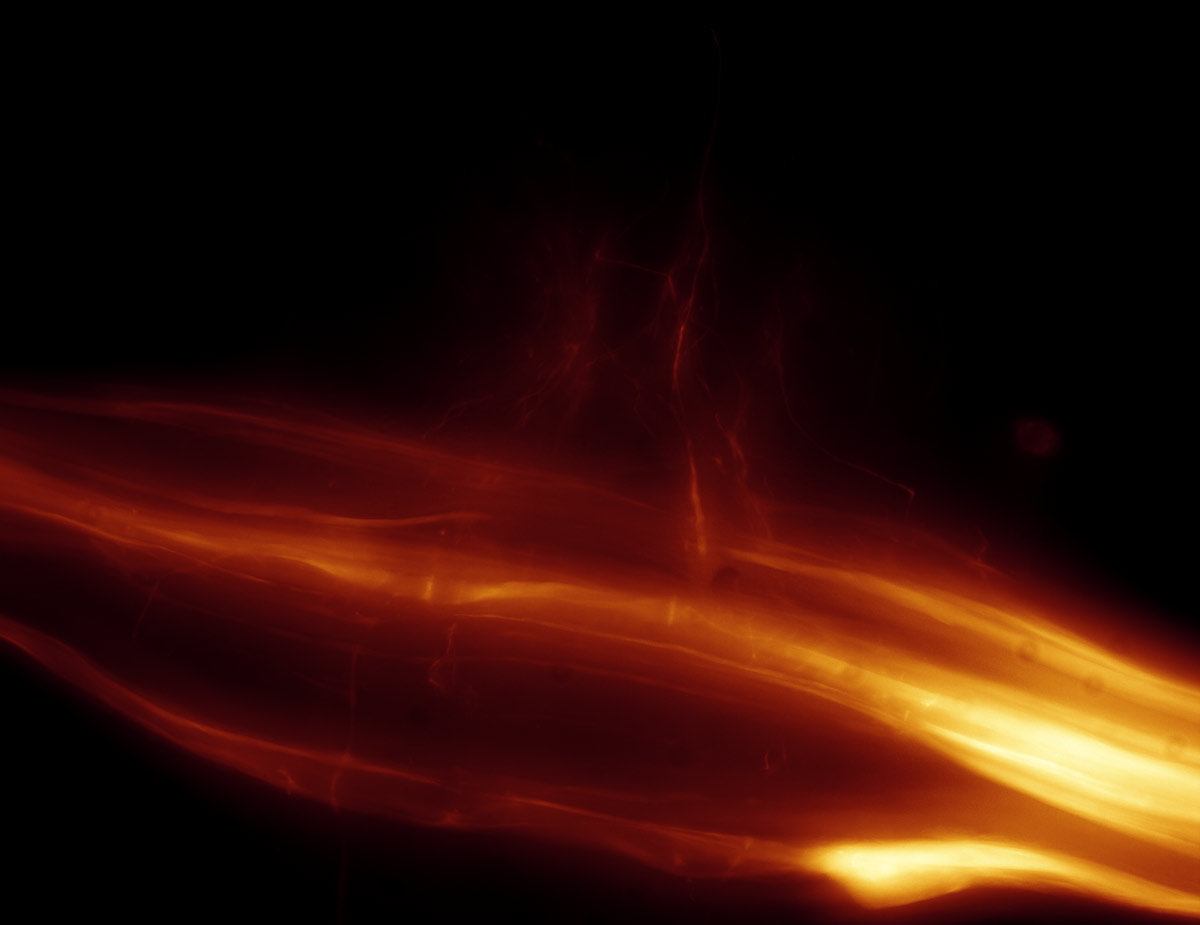
従来の蛍光画像THUNDER画像

バッタの神経節、従来の蛍光画像(左)、THUNDER 像(右)。厚さ:110µm、データ量:376 MB。Computational Clearing による画像取得時間:3 秒

実感できるスピード - THUNDER Live

実感できるスピード

Show details

実感できるスピード

THUNDER で用いられる Computational Clearing はリアルタイムに作動します。その計算処理は画像取得と同時に開始されます。THUNDER は画像取得のフローに直接組み込まれ、イメージングに用いられる光学的なパラメータとソフトウェア上の設定をすべて採用します。

この「光学との融合」によって、THUNDER による画像は驚くほど高速に画面表示され、閲覧や評価が可能となります。従来のように終了まで長時間も待つ必要がありません。

THUNDER のスピードは見て感じることのできるものです。

優れた結果を容易に達成

THUNDER はワンクリック・オペレーションにより、迅速に動作します。 システムの較正やハードウェアのコンポーネントの調整は必要ありません。Computational Clearing の手法を選択するだけで、エキスパートレベルの結果がわずかな時間で得られます。

複数のサンプルで実験を繰り返すことも容易です。THUNDER のイメージングパラメータも簡単に保存、素早く読み出しもできます。

「大型サンプルでも 2 分以内に驚くほど鮮明な画像を提供する THUNDER はタイムラプス試験に有効です」
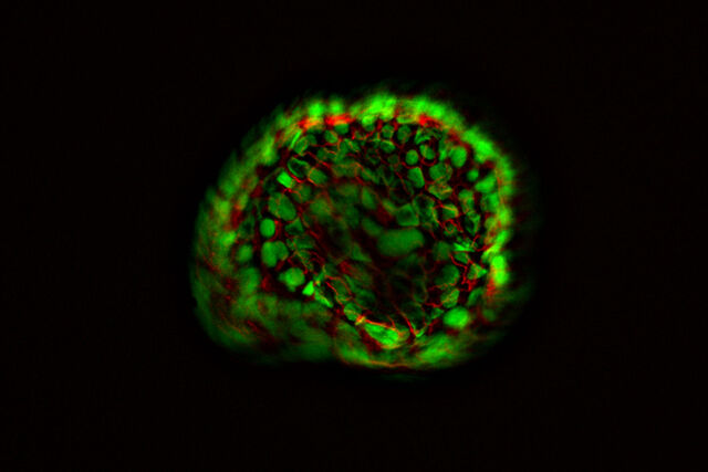
Almary Guerra 博士、マックス・プランク心肺研究所(ドイツ、バート・ナウハイム)

Computational Clearing

Computational Clearing は、観察しようとする部位のサイズを考慮しながら、シグナルとバックグラウンドとを効果的に識別します。これにより、従来は抜け落ちていたサンプルの微弱なシグナルも可視化できます。1 枚の平面像を取得するだけで、驚くべき結果が画面に表示されます。

メソッドの選択肢には、ライカの共焦点顕微鏡システムでおなじみのデシジョンマスクを用いたデコンボリューションとの組み合わせもご用意しています。完全に自動化されたこのメソッドは、ユーザーによる手入力なしで計算処理します。これにより極めて高速高品質の画像が生成されます。

信頼性ある結果で次のステップへ

Computational Clearing を 1 回起動するだけで、ボケのない画像が得られます。実験が単純であるか複雑であるかには関わりなく、RAW データは常に保存され、データの見直しにも最適です。

この高精細の THUNDER 像を使って、細胞核のカウント、核内ドットのカウント、細胞のトラッキング等、様々な解析を進めることができます。

「ボケ」のないクリアな画像

THUNDER システムはライカ独自の新技術「Computational Clearing」によって、広視野観察に見られるフォーカスアウトした部分のボケ情報を取り除きます。このため THUNDER を利用すれば、WideField システムの速度と感度を維持しつつ同時に厚みのある立体的なサンプルでも高品質の 3D 画像を得ることができます。

Scroll to top