Infinity TIRF
アクセサリー
製品紹介
Home
Leica Microsystems
Infinity TIRF DMi8 S モジュール
隠されているものを確認
最新の記事を読む
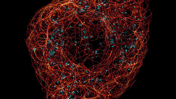
A Guide to Super-Resolution
Find out more about Leica super-resolution microscopy solutions and how they can empower you to visualize in fine detail subcellular structures and dynamics.
ウイルス学
ウイルス研究のためのイメージングと試料作製ソリューション
Universal PAINT – Dynamic Super-Resolution Microscopy
Super-resolution microscopy techniques have revolutionized biology for the last ten years. With their help cellular components can now be visualized at the size of a protein. Nevertheless, imaging…
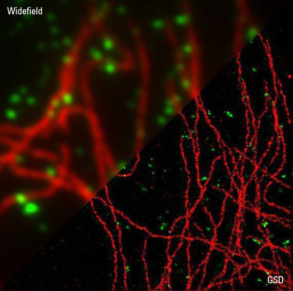
Sample Preparation for GSDIM Localization Microscopy – Protocols and Tips
The widefield super-resolution technique GSDIM (Ground State Depletion followed by individual molecule return) is a localization microscopy technique that is capable of resolving details as small as…
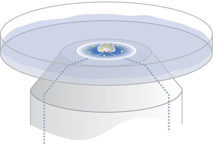
Controlling the TIRF Penetration Depth is Mandatory for Reproducible Results
The main feature of total internal reflection fluorescence (TIRF) microscopy is the employment of an evanescent wave for the excitation of fluorophores instead of using direct light. A property of the…
Total Internal Reflection Fluorescence (TIRF) Microscopy
Total internal reflection fluorescence (TIRF) is a special technique in fluorescence microscopy developed by Daniel Axelrod at the University of Michigan, Ann Arbor in the early 1980s. TIRF microscopy…
Super-Resolution GSDIM Microscopy
The nanoscopic technique GSDIM (ground state depletion microscopy followed by individual molecule return) provides a detailed image of the spatial arrangement of proteins and other biomolecules within…
応用分野
細胞生物学
ヒトの健康と病気を細胞ベースで理解することを目的として研究を行う場合、関心のある細胞の構造および分子の詳細から対象の細胞を研究することが重要です。 その結果、細胞生物学における顕微鏡はかってないほどに重要なツールとなり、構造環境内で試料を詳細に調査したり、細胞内小器官や高分子を分析したりすることができます。 細胞生物学イメージングは、さまざまな光電子相関顕微鏡を使用して行われます。…
