DM IL LED
倒立顕微鏡
光学顕微鏡
製品紹介
Home
Leica Microsystems
DM IL LED 研究用倒立顕微鏡
最新の記事を読む
Phase Contrast and Microscopy
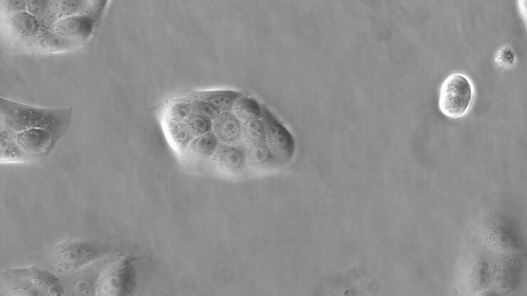
This article explains phase contrast, an optical microscopy technique, which reveals fine details of unstained, transparent specimens that are difficult to see with common brightfield illumination.
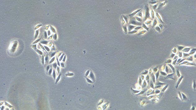
How to do a Proper Cell Culture Quick Check
In order to successfully work with mammalian cell lines, they must be grown under controlled conditions and require their own specific growth medium. In addition, to guarantee consistency their growth…
Microscopy in Virology
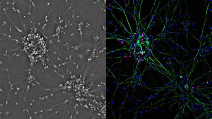
The coronavirus SARS-CoV-2, causing the Covid-19 disease effects our world in all aspects. Research to find immunization and treatment methods, in other words to fight this virus, gained highest…
Introduction to Mammalian Cell Culture
Mammalian cell culture is one of the basic pillars of life sciences. Without the ability to grow cells in the lab, the fast progress in disciplines like cell biology, immunology, or cancer research…
Introduction to Widefield Microscopy
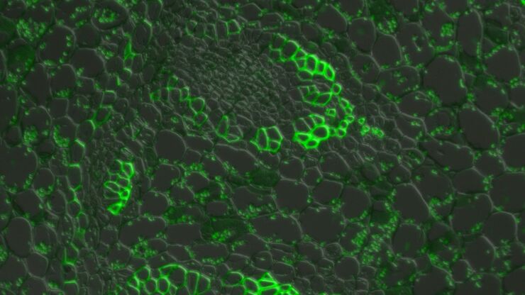
This article gives an introduction to widefield microscopy, one of the most basic and commonly used microscopy techniques. It also shows the basic differences between widefield and confocal…
応用分野
細胞培養
ラボ環境での細胞培養は、細胞生物学、がん研究、発生生物学をはじめ、ライフサイエンスに関するすべての分野、そして薬学の研究の基本です。ライカが研究室内での細胞培養をどのようにサポートしているか、こちらでご覧いただけます。


