
Filter articles
标签
Story Type
Products
Loading...

How to Save Time and Samples by Automated Ultramicrotomy
This article describes how 3D micro-CT data of a resin-embedded electron microscopy sample can be used to trim the specimen down to a defined target plane prior to sectioning. The interactive and…
Loading...
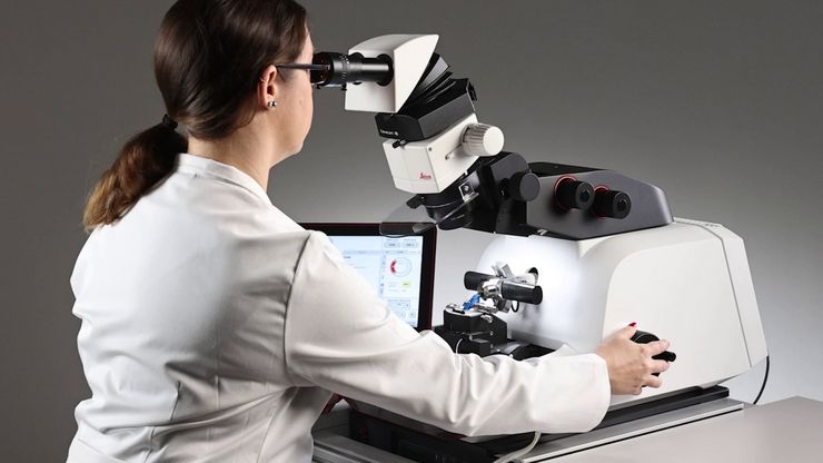
Essential Guide to Ultramicrotomy
When studying samples, to visualize their fine structure with nanometer scale resolution, most often electron microscopy is used. There are 2 types: scanning electron microscopy (SEM) which images the…
Loading...
 applied. Image courtesy of Samuel East, Uncommon Bio.")
Designing the Future with Novel and Scalable Stem Cell Culture
Visionary biotech start-up Uncommon Bio is tackling one of the world’s biggest health challenges: food sustainability. In this webinar, Stem Cell Scientist Samuel East shows how they make stem cell…
Loading...
, microglia (TMEM119, IBA1), and Alzheimer’s-associated markers (β-amyloid and p-Tau217).")
Explore Alzheimer's Spatial Proteome with Big Data
Alzheimer's disease, a genetic and sporadic neurodegenerative condition, leads to cognitive decline in mid to late life, marked by β-amyloid plaques and tau tangles. With limited treatment options,…
Loading...

Automotive Part Verification and Development according to Specifications
Automotive part verification during the development and production of parts and components by suppliers or manufacturers is important for ensuring that specifications are met. Specifications are…
Loading...

Mica: A Game-changer for Collaborative Research at Imperial College London
This interview highlights the transformative impact of Mica at Imperial College London. Scientists explain how Mica has been a game-changer, expanding research possibilities and facilitating…
Loading...

From Bench to Beam: A Complete Correlative Cryo Light Microscopy Workflow
In the webinar entitled "A Multimodal Vitreous Crusade, a Cryo Correlative Workflow from Bench to Beam" a team of experts discusses the exciting world of correlative workflows for structural biology…
Loading...
 which was imaged with extended depth of field (EDOF) using digital microscopy.")
顕微鏡を知る:被写界深度
顕微鏡において被写界深度は、凹凸の変化が⼤きい構造を持つ試料をピントがあったシャープに観察・撮像するために重要なパラメータです。被写界深度は、開⼝数、解像度、倍率の相関関係によって決定され、解像度とパラメータは反⽐例の関係にあります。被写界深度と解像度のバランスが最適になるように調整することができる顕微鏡もあります。
Loading...

がん研究
がんは、成長調節における欠損細胞によって引き起こされる複雑な異質性疾患です。 細胞または細胞群内の遺伝的および後成的変化が通常の機能を妨げ、自律的、非制御の細胞成長と増殖を引き起こします。
