
Filter articles
标签
Products
Loading...
レーザーマイクロダイセクション
小動物などの解剖を行うとき、顕微鏡の接眼レンズを何時間ものぞくことがあります。 ライカ マイクロシステムズでは、さまざまな顕微鏡と幅広い解剖顕微鏡部品やアクセサリーから選ぶことができるため、ニーズに最適な顕微鏡ソリューションを見つけることができます。
Loading...
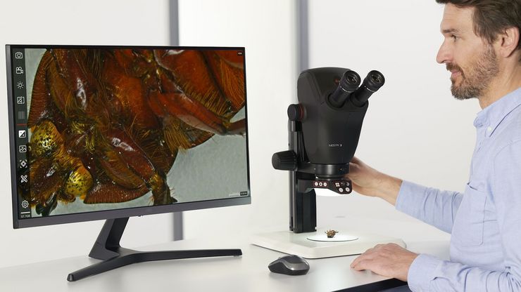
 stereo microscope for a task like surgery.")
Rodent and Small-Animal Surgery
Learn how you can perform rodent (mouse, rat, hamster) and small-animal surgery efficiently with a microscope for developmental biology and medical research applications by reading this article.
Loading...

Imaging and Analyzing Zebrafish, Medaka, and Xenopus
Discover how to image and analyze zebrafish, medaka, and Xenopus frog model organisms efficiently with a microscope for developmental biology applications from this article.
Loading...

Investigating Fruit Flies (Drosophila melanogaster)
Learn how to image and investigate Drosophila fruit fly model organisms efficiently with a microscope for developmental biology applications from this article.
Loading...

The History of Stereo Microscopy
This article gives an overview on the history of stereo microscopes. The development and evolution from handcrafted instruments (late 16th to mid-18th century) to mass produced ones the last 150…
