
Filter articles
标签
Story Type
Products
Loading...

Multi-Color Caspase 3/7 Assays with Mica
Caspases are involved in apoptosis and can be utilized to determine if cells are undergoing this programmed cell death pathway in so-called caspase assays. These assays can be run by e.g. flow…
Loading...

Find Relevant Specimen Details from Overviews
Switch from searching image by image to seeing the full overview of samples quickly and identifying the important specimen details instantly with confocal microscopy. Use that knowledge to set up…
Loading...

How to Successfully Perform Live-Cell CLEM
The Leica Nano workflow provides a streamlined live-cell CLEM solution for getting insight bout structural changes of cellular components over time. Besides the technical handling described in the…
Loading...

Consumables for Laser Microdissection
There are many different types of consumables for laser microdissection (LMD) systems. They cover a wide range of applications from basic to highly specialized, enabling scientists to choose their own…
Loading...
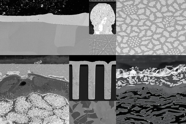
Introduction to Ion Beam Etching with the EM TIC 3X
In this article you can learn how to optimize the preparation quality of your samples by using the ion beam etching method with the EM TIC 3X ion beam milling machine. A short introduction of the…
Loading...
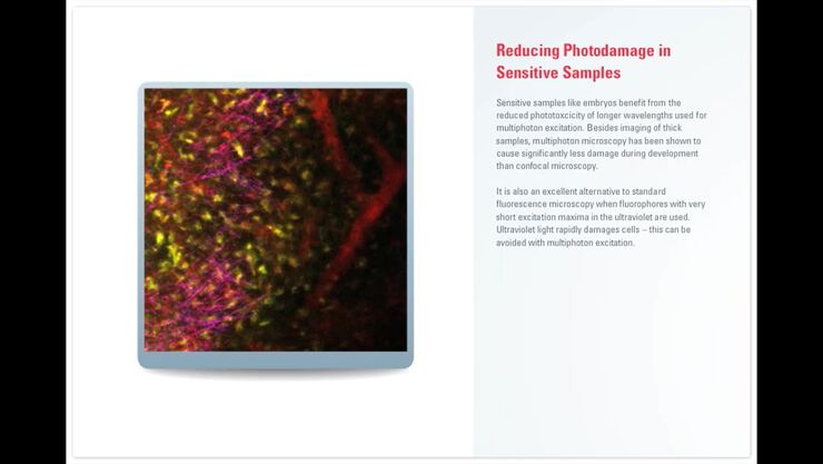
Principles of Multiphoton Microscopy for Deep Tissue Imaging
This tutorial explains the principles of multiphoton microscopy for deep tissue imaging. Multiphoton microscopy uses excitation wavelengths in the infrared taking advantage of the reduced scattering…
Loading...
![3D glomeruli in a portion of an ECi-cleared kidney scanned by light sheet microscopy. Courtesy of Prof. Norbert Gretz, Medical Faculty Mannheim, University of Heidelberg [1].](/fileadmin/_processed_/d/d/csm_DLS-Sample-Preparation-Intr_915e0fd7c2.jpg "3D glomeruli in a portion of an ECi-cleared kidney scanned by light sheet microscopy. Courtesy of Prof. Norbert Gretz, Medical Faculty Mannheim, University of Heidelberg [1].")
Using Mounting Frames for Light Sheet Sample Preparation
Sample handling is an important topic in the context of Light Sheet Microscopy. The TCS SP8 DLS integrates Light Sheet technology into an inverted confocal platform and can hence make use of general…
Loading...

Using a Rotation Device for Light Sheet Sample Mounting
The TCS SP8 DLS from Leica Microsystems is an innovative concept to integrate the Light Sheet Microscopy technology into the confocal microscope. Due to its unique optical architecture samples can be…
Loading...
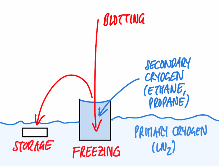
Immersion Freezing for Cryo-Transmission Electron Microscopy: Fundamentals
The high vacuum required in a transmission electron microscope (TEM) greatly impairs the ability to study specimens naturally occurring in an aqueous phase: exposing "wet" specimens to a pressure…
