
Filter articles
标签
Story Type
Products
Loading...
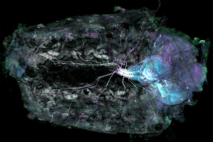
Understanding Motor Sequence Generation Across Spatiotemporal Scales
We have developed a microscopy-based pipeline to characterize a developmentally critical behavior at the pupal stage of development, called the ecdysis sequence. We study brain-wide neuronal activity…
Loading...
Investigating Synapses in Brain Slices with Enhanced Functional Electron Microscopy
A fundamental question of neuroscience is: what is the relationship between structural and functional properties of synapses? Over the last few decades, electrophysiology has shed light on synaptic…
Loading...
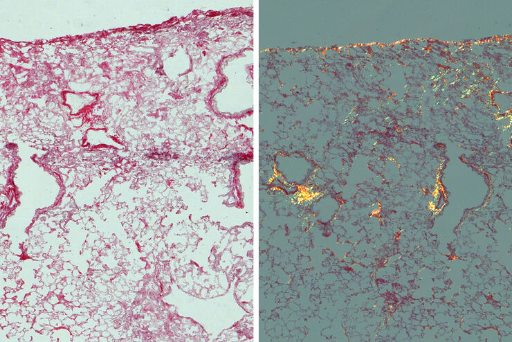
Studying Pulmonary Fibrosis
The results shown in this article demonstrate that fibrotic and non-fibrotic regions of collagen present in mouse lung tissue can be distinguished better with polarized light compared to brightfield.…
Loading...

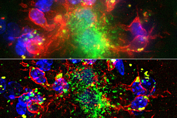
Image Gallery: THUNDER Imager
To help you answer important scientific questions, THUNDER Imagers eliminate the out-of-focus blur that clouds the view of thick samples when using camera-based fluorescence microscopes. They achieve…
Loading...

研究におけるモデル生物
モデル生物とは、特定の生物学的プロセスを研究するために研究者が使用する生物種です。 モデル生物は、人間と似た遺伝的特徴を持ち、遺伝子学、発生生物学、神経科学などの研究分野で一般的に使用されています。 通常、モデル生物は実験環境での維持や繁殖が容易であること、生殖サイクルが短いこと、または、特定の形質や病気を研究するために突然変異体を生成する能力を持つことで選ばれます。
Loading...

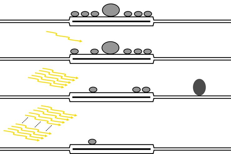
An Introduction to Computational Clearing
Many software packages include background subtraction algorithms to enhance the contrast of features in the image by reducing background noise. The most common methods used to remove background noise…
Loading...
Computational Clearing - Enhance 3D Specimen Imaging
This webinar is designed to clarify crucial specifications that contribute to THUNDER Imagers' transformative visualization of 3D samples and improvements within a researcher's imaging-related…
Loading...
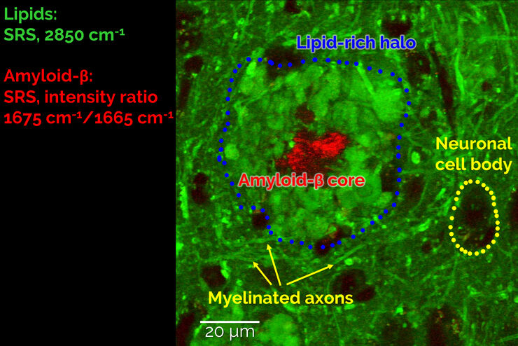
Stimulated Raman Scattering Microscopy Probes Neurodegenerative Disease
Despite decades of research, the molecular mechanisms underlying some of the most severe neurodegenerative diseases, such as Alzheimer’s or Parkinson’s, remain poorly understood. The progression of…
Loading...

Zebrafish Brain - Whole Organ Imaging at High Resolution
Structural information is key when one seeks to understand complex biological systems, and one of the most complex biological structures is the vertebrate central nervous system. To image a complete…
