
Filter articles
标签
Story Type
Products
Loading...

Introduction to 21 CFR Part 11 and Related Regulations
This article provides an overview of regulations and guidelines for electronic records (data entry, storage, signatures, and approvals) used in the USA (21 CFR Part 11), EU (GMP Annex 11), and China…
Loading...

Physiology Image Gallery
Physiology is about the processes and functions within a living organism. Research in physiology focuses on the activities and functions of an organism’s organs, tissues, or cells, including the…
Loading...

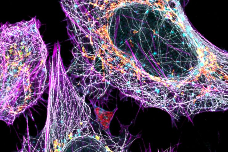
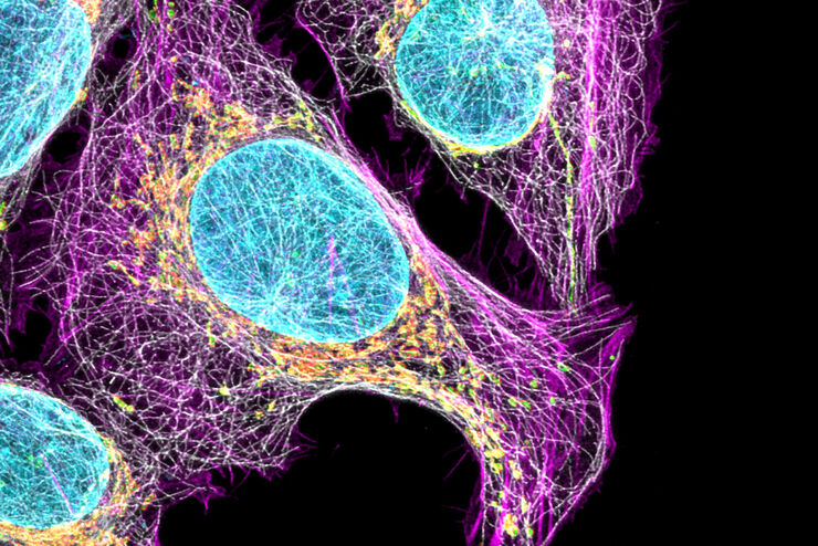
Live Cell Imaging Gallery
Live cell microscopy techniques are fundamental to get a better understanding of cellular and molecular function. Today, widefield microscopy is the most common technique used to visualize cell…
Loading...

Tissue Image Gallery
Visual analysis of animal and human tissues is critical to understand complex diseases such as cancer or neurodegeneration. From basic immunohistochemistry to intravital imaging, confocal microscopy…
Loading...
 and astrocytes (green) in a cortical spheroid derived from human induced pluripotent stem cells.")
Neuroscience Images
Neuroscience commonly uses microscopy to study the nervous system’s function and understand neurodegenerative diseases.
Loading...
Multicolor Image Gallery
Fluorescence multicolor microscopy, which is one aspect of multiplex imaging, allows for the observation and analysis of multiple elements within the same sample – each tagged with a different…
Loading...

Cancer Research Image Gallery
Fluorescence microscopy allows the study of changes occurring in tissue and cells during cancer development and progression. Techniques such as live cell imaging are critical to understand cancer…
Loading...
, TL DIC")
微分干渉(DIC)顕微鏡
微分干渉(DIC)顕微鏡は、光源と集光レンズの間に偏光フィルターとウォラストンプリズムを配置した広視野顕微鏡で、対物レンズとカメラセンサーや接眼レンズの間にも偏光フィルターとウォラストンプリズムを配置しています。
Loading...
Cell Biology Image Gallery
Cell biology studies the structure, function and behavior of cells, including cell metabolism, cell cycle, and cell signaling. Fluorescence microscopes are an integral part of a cell biologist…
