
Filter articles
标签
Products
Loading...

What is the FusionOptics Technology?
Leica stereo microscopes with FusionOptics provide optimal 3D perception. The brain merges two images, one with large depth of field and the other with high resolution, into one 3D image.
Loading...

Microscope Ergonomics
This article explains microscope ergonomics and how it helps users work in comfort, enabling consistency and efficiency. Learn how to set up the workplace to keep good posture when using a microscope.
Loading...

Digital Inspection Microscope for Industrial Applications
Factors users should consider before choosing a digital inspection microscope for industrial applications, including quality control (QC), failure analysis (FA), and R&D, are described in this…
Loading...
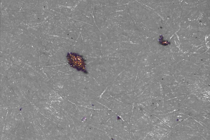
 taken with a ring light (RL) and near vertical illumination (NVI).")
Microscope Illumination for Industrial Applications
Inspection microscope users can obtain information from this article which helps them choose the optimal microscope illumination or lighting system for inspection of parts or components.
Loading...

Alternative Fuels and Why Sustainable Solutions are Important
This free on-demand webinar is about the role of alternative fuel vehicles and why sustainable solutions are of increasing importance to the automotive industry.
Loading...

Technical Cleanliness in the Automotive Industry for Electromobility
This free on-demand webinar covers the increasing focus on technical cleanliness in the automotive industry for electromobility and the VDA 19.1 revision.
Loading...

3 Factors Determine the Damage Potential of Particles
This article discusses the 3 factors for determining the potential of a particle to cause damage to parts and components in the automotive and electronic industry. These factors include the…
Loading...
Factors to Consider for a Cleanliness Analysis Solution
Choosing the right cleanliness analysis solution is important for optimal quality control. This article discusses the important factors that should be taken into account to find the solution that best…
Loading...

H&E Staining in Microscopy
If we consider the role of microscopy in pathologists’ daily routines, we often think of the diagnosis. While microscopes indeed play a crucial role at this stage of the pathology lab workflow, they…
