
Filter articles
标签
Products
Loading...

Optimizing THUNDER Platform for High-Content Slide Scanning
With rising demand for full-tissue imaging and the need for FL signal quantitation in diverse biological specimens, the limits on HC imaging technology are tested, while user trainability and…
Loading...
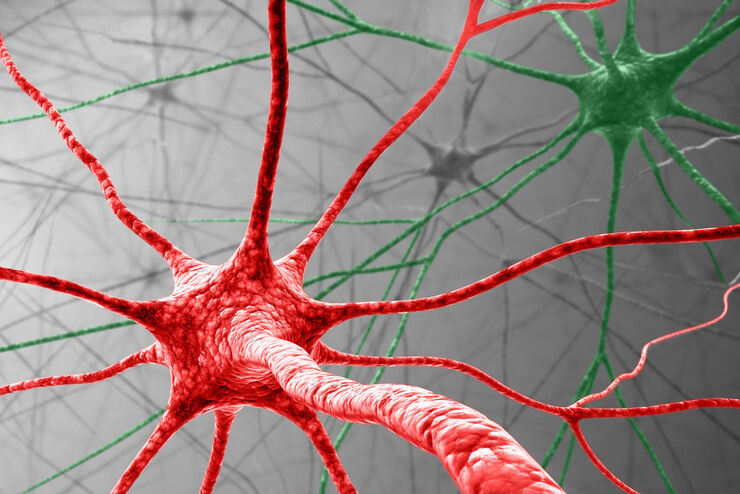
Visualizing Protein Degradation and Aggregation in the Living Cell
Our guest speaker, Prof Dr Eric Reits, presents his work on neurodegenerative disorders. Reits’ group are experts on the subject of Huntington’s disease and work towards identifying leads for…
Loading...
Capture life as it happens
With the Leica Nano Workflow, searching for the needle in the haystack is a thing of the past. Take advantage of correlative light and electron microscopy to identify directly the right cell at the…
Loading...
Life Beyond the Pixels: Deep Learning Methods for Single Cell Analysis
Our guest speaker Prof Dr Peter Horvath presents his work on single cell-based large-scale microscopy experiments. This novel targeting approach includes the use of machine learning models and…
Loading...

Tissue Image Gallery
Visual analysis of animal and human tissues is critical to understand complex diseases such as cancer or neurodegeneration. From basic immunohistochemistry to intravital imaging, confocal microscopy…
Loading...
, TL DIC")
微分干渉(DIC)顕微鏡
微分干渉(DIC)顕微鏡は、光源と集光レンズの間に偏光フィルターとウォラストンプリズムを配置した広視野顕微鏡で、対物レンズとカメラセンサーや接眼レンズの間にも偏光フィルターとウォラストンプリズムを配置しています。
Loading...

Dissecting Proteomic Heterogeneity of the Tumor Microenvironment
This lecture will highlight cutting edge applications in applying laser microdissection and microscaled quantitative proteomics and phosphoproteomics to uncover exquisite intra- and inter-tumor…

Loading...
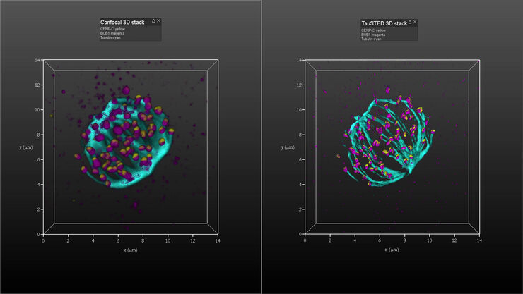
Kinetochore Assembly during Mitosis with TauSTED on 3D
Three-dimensional organization of the mitotic spindle together with the distribution of CENP-C and BUB1 based on TauSTED with multiple STED lines (592, 660 and 775 nm) can provide insights…
