
Filter articles
标签
Story Type
Products
Loading...

Going Beyond Deconvolution
Widefield fluorescence microscopy is often used to visualize structures in life science specimens and obtain useful information. With the use of fluorescent proteins or dyes, discrete specimen…
Loading...
 taken with a ring light (RL) and near vertical illumination (NVI).")
Microscope Illumination for Industrial Applications
Inspection microscope users can obtain information from this article which helps them choose the optimal microscope illumination or lighting system for inspection of parts or components.
Loading...
 cells taken with phase contrast.")
Phase Contrast and Microscopy
This article explains phase contrast, an optical microscopy technique, which reveals fine details of unstained, transparent specimens that are difficult to see with common brightfield illumination.
Loading...

Surgical Management of High-Grade Gliomas
Learn about the surgical management of high-grade gliomas and how to expand the extent of resection intra-operatively using tools such as 5-ALA fluorescence.
Loading...

How to Radically Simplify Workflows in Your Imaging Facility
VIDEO ON DEMAND - How to radically simplify imaging workflows and generate meaningful results with less time and effort using a highly automated microscope that unites widefield and confocal imaging.
Loading...
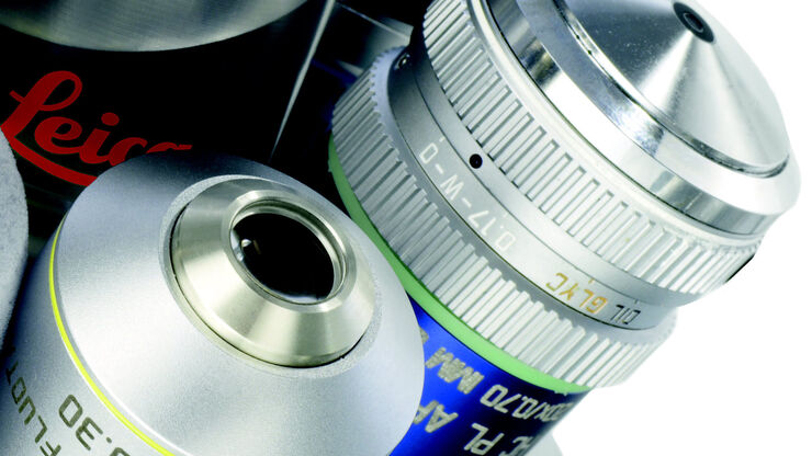
Immersion Objectives
How an immersion objective, which has a liquid medium between it and the specimen being observed, helps increase the numerical aperture and microscope resolution is explained in this article.
Loading...

Advancing Surgery in Pediatric Brain Tumors
Learn how fluorescence-guided surgery supports pediatric brain tumor resection and improves precision through literature review and clinical cases.
Loading...

How to Determine Cell Confluency with a Digital Microscope
This article shows how to measure cell confluency in an easy and consistent way with Mateo TL, increasing confidence in downstream experiments.
Loading...

How to do a Proper Cell Culture Quick Check
In order to successfully work with mammalian cell lines, they must be grown under controlled conditions and require their own specific growth medium. In addition, to guarantee consistency their growth…
