is mobile? false
Science Lab
Science Lab
ライカマイクロシステムズのナレッジポータルでは、顕微鏡の基礎から最先端技術まで、幅広い情報を提供しています。初心者から熟練者、研究者、医師の皆様まで、日々の研究や実験に役立つ内容となっております。チュートリアルやアプリケーションノートを活用し、学びながら探究心を刺激してください。さらに、コミュニティに参加することで、知見を共有し、新たな発見へとつなげましょう。お気軽に参加いただき、互いの専門知識を深め合う場としてご活用ください。
ナレッジポータル Show subnavigation


アプリケーションの種類 Show subnavigation


Filter articles
标签
Story Type
Products
Loading...

神経科学研究
神経変性疾患の理解向上に取り組んでいる、もしくは神経系の機能を研究をしていますか? ライカマイクロシステムズのイメージングソリューションによってブレイクスルーを起こす方法をご覧ください。
Loading...

Mapping Tumor Immune Landscape with AI-Powered Spatial Proteomics
Spatial mapping of untreated tumors provides an overview of the tumor immune architecture, useful for understanding therapeutic responses. Immunocompetent murine models are essential for identifying…

Loading...
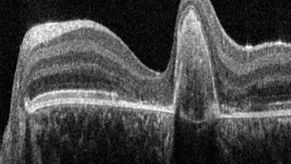
A Guide to OCT
Leica Optical Coherence Tomography (OCT) systems support ophthalmologists, ophthalmic surgeons, and researchers with easy-to-use, high-quality imaging technology.

Loading...

ゼブラフィッシュを用いた研究
スクリーニング、ソーティング、マニピュレーションおよびイメージングを通じて最良の結果を得るためには、細部や構造を観察して、研究の次の段階に向けて正しい判断を下す必要があります。
優れた光学系と高解像度で定評のあるライカの実体顕微鏡と透過照明スタンドは、世界中の研究者から支持されています。
Loading...

Aneurysm Clipping: Assessing Perforators in Real-Time with AR Fluorescence
This article covers two aneurysm clipping cases highlighting the clinical benefits of GLOW800 Augmented Reality Fluorescence application in neurosurgery, based on insights from Prof. Tohru Mizutani,…
Loading...

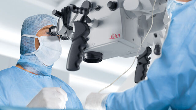
How does Real-Time OCT Imaging Impact Precision in Corneal Surgery?
Corneal surgery is a highly specialized field. It requires great surgical precision to overcome challenges such as visualizing clearly the full anterior chamber, performing Descemet membrane peeling…
Loading...

How to Achieve Brain Tissue Resection with GLOW400 AR
Intraoperative MRI is one form of real-time intraoperative visualization, but if more in-depth visualization to identify a tumor during surgery is wanted, intraoperative fluorescence diagnostics is…
