
Filter articles
标签
Story Type
Products
Loading...

The Principles of White Light Laser Confocal Microscopy
The perfect light source for confocal microscopes in biomedical applications has sufficient intensity, tunable color and is pulsed for use in lifetime fluorescence. Furthermore, it should offer means…
Loading...

Optical Microscopes – Some Basics
The optical microscope has been a standard tool in life science as well as material science for more than one and a half centuries now. To use this tool economically and effectively, it helps a lot to…
Loading...
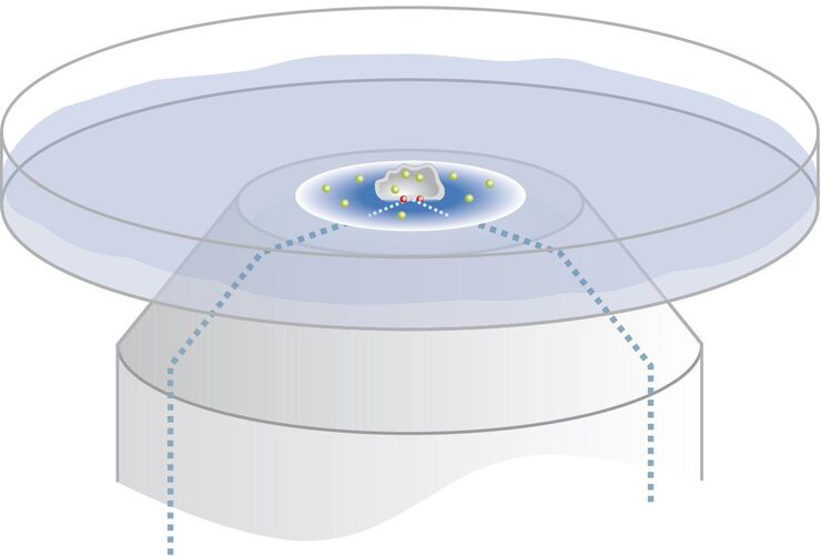
Controlling the TIRF Penetration Depth is Mandatory for Reproducible Results
The main feature of total internal reflection fluorescence (TIRF) microscopy is the employment of an evanescent wave for the excitation of fluorophores instead of using direct light. A property of the…
Loading...
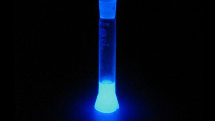
Basic Principles of Luminescence
There are a lot of light-emitting processes occurring in nature. Luminescence is an umbrella term for those kinds of events where light emission is not the result of high temperatures. This article…
Loading...
 inoculated with cowpea mosaic virus (CPMV) containing the GFP-gene inserted between the movement protein (MP) and the capsid proteins (CPs) in the viral RNA 2")
Introduction to Live-Cell Imaging
The understanding of complex and fast cellular dynamics is an important step to get insight into biological processes. Therefore, today’s life science research more and more demands studying…
Loading...

Total Internal Reflection Fluorescence (TIRF) Microscopy
Total internal reflection fluorescence (TIRF) is a special technique in fluorescence microscopy developed by Daniel Axelrod at the University of Michigan, Ann Arbor in the early 1980s. TIRF microscopy…
Loading...

Applications of TIRF Microscopy in Life Science Research
The special feature of TIRF microscopy is the employment of an evanescent field for fluorophore excitation. Unlike standard widefield fluorescence illumination procedures with arc lamps, LEDs or…
Loading...

Fluorescent Proteins - From the Beginnings to the Nobel Prize
Fluorescent proteins are the fundament of recent fluorescence microscopy and its modern applications. Their discovery and consequent development was one of the most exciting innovations for life…
Loading...

Ratiometric Imaging
Many fundamental functions of a cell strongly depend on delicate, but nevertheless dynamic balances of ions (e.g. calcium, magnesium), voltage potentials and pH between the cell’s cytosol and the…
