ウイルス学

イメージングと試料作製のためのライカソリューションは、ウイルスの侵入、融合、ゲノム統合、ウイルス複製、ウイルス集合、ウイルス出芽の調査に有効です。

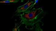
 Influenza_lung_porcine_ICC_3-2.jpg

ウイルス感染メカニズムを理解し、その疾患の治療法を開発するためのウイルス感染組織と細胞の研究は、ヒトの健康に大きく寄与します。 イメージングと試料作製のためのライカソリューションは、ウイルスの侵入、融合、ゲノム統合、ウイルス複製、ウイルス集合、ウイルス出芽の調査に有効です。 また、適切に準備された試料の分析から得られるデータは、関連する細胞メカニズムと免疫反応の研究にとって重要です。 ウイルス研究の最終的な目標は、介入戦略の開発です。

ウイルスは生体検査から単一ウイルス粒子レベルまで研究されます。 最も広く研究されているウイルスには、インフルエンザ、コロナウイルス(COVID-19の原因となるSARS-CoV-2等)、ヘルペス、肝炎、デング熱、ジカ、HIV、狂犬病、エボラ等がありますが、さらに多くのウイルスがあります。

ウイルス感染過程の宿主側

ウイルス研究ではモデル生物が用いられることもありますが、ヒトの宿主の形態をうまく模倣することはできません。 このため、研究者はヒト細胞、生体検査、細胞培養研究に頼ることが多くなります。

生体外感染モデルは組織、生体検査、動物モデルから生成することができます。 ライカ実体顕微鏡はこのようなモデル研究に効果的です。

ライカのワイドフィールド顕微鏡を使用することで、細胞培養、スフェロイド、オルガノイドをモニタリングすることが可能です。

THUNDERイメージャーといったライカの蛍光顕微鏡を活用して、感染モデル、2Dまたは3D細胞培養の初期研究をさらに深めることができます。

特定の細胞を周囲組織から分離する、ライカマイクロシステムズのレーザーマイクロダイセクションによって、より確かな遺伝子分析を実施できます。

ライカソリューションのウイルス研究アプリケーションはこちらのアプリケーションノートでご覧いただけます。

Related articles

Microscopy in Virology

The coronavirus SARS-CoV-2, causing the Covid-19 disease effects our world in all aspects. Research to find immunization and treatment methods, in other words to fight this virus, gained highest…
Image source: Adobe Stock

How to Sanitize a Microscope

Due to the current coronavirus pandemic, there are a lot of questions regarding decontamination methods of microscopes for safe usage. This informative article summarizes general decontamination…

ウイルス研究のための高分解能蛍光イメージング

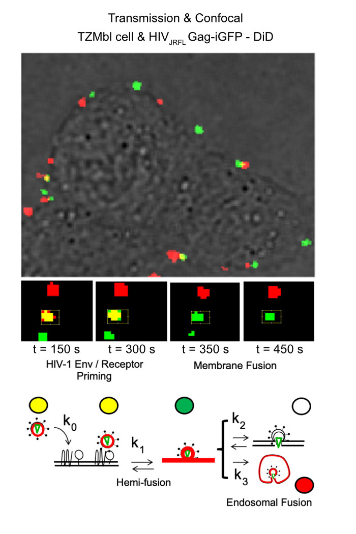
原因となるウイルスや病原体を追跡することを可能にする高分解能顕微鏡観察は、感染症研究にとって有用です。 細胞やウイルス成分の蛍光標識と組み合わせることで、感染過程の複数の側面を研究することができます。

このため、超高分解能を得る共焦点レーザー顕微鏡と全反射照明蛍光(TIRF)顕微鏡は、共にウイルス研究に最適なツールです。

多光子とライトシートによる顕微鏡観察といった高度な顕微鏡技術は、組織切片、動物モデル、オルガノイド等の3D感染モデルといった異なる状況下で発生する感染事例にアクセスすることを可能にします。

STELLARIS共焦点プラットフォームとTIRF ソリューションを含むライカの顕微鏡は、ウイルス研究に関連した3D試料の研究にとって多くの利点を提供します。

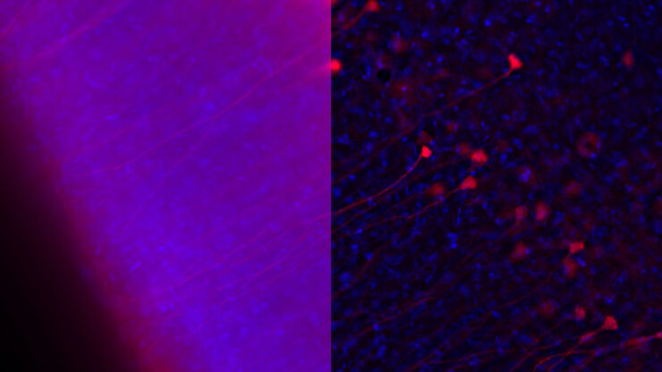
画像および図表出典:Coomer et al. PLOS Pathogen (2020) 16(2): e1008359, DOI: 10.1371/journal.ppat.1008359.
Related articles

Molecular structure of the green fluorescent protein (GFP)

Introduction to Fluorescent Proteins

Overview of fluorescent proteins (FPs) from, red (RFP) to green (GFP) and blue (BFP), with a table showing their relevant spectral characteristics.
Cellular microtubule network in a fibroblast cell.

How to Prepare your Specimen for Immunofluorescence Microscopy

Immunofluorescence (IF) is a powerful method for visualizing intracellular processes, conditions and structures. IF preparations can be analyzed by various microscopy techniques (e.g. CLSM,…
Image of immunofluorescently labelled cells where mitochondria are indicated with red, nuclei with blue, and actin with green.

Studying Virus Replication with Fluorescence Microscopy

The results from research on SARS-CoV-2 virus replication kinetics, adaption capabilities, and cytopathology in Vero E6 cells, done with the help of fluorescence microscopy, are described in this…
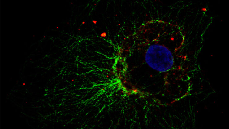
Influenca in lung epithelial cells (porcine) - THUNDER Imager 3D Cell Culture Influenca virus – red, cilia – green, Nuclei – blue.

How Can Immunofluorescence Aid Virology Research?

Modern virology research has become as crucial now as ever before due to the global COVID-19 pandemic. There are many powerful technologies and assays that virologists can apply to their research into…

ウイルスの超微細構造

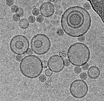
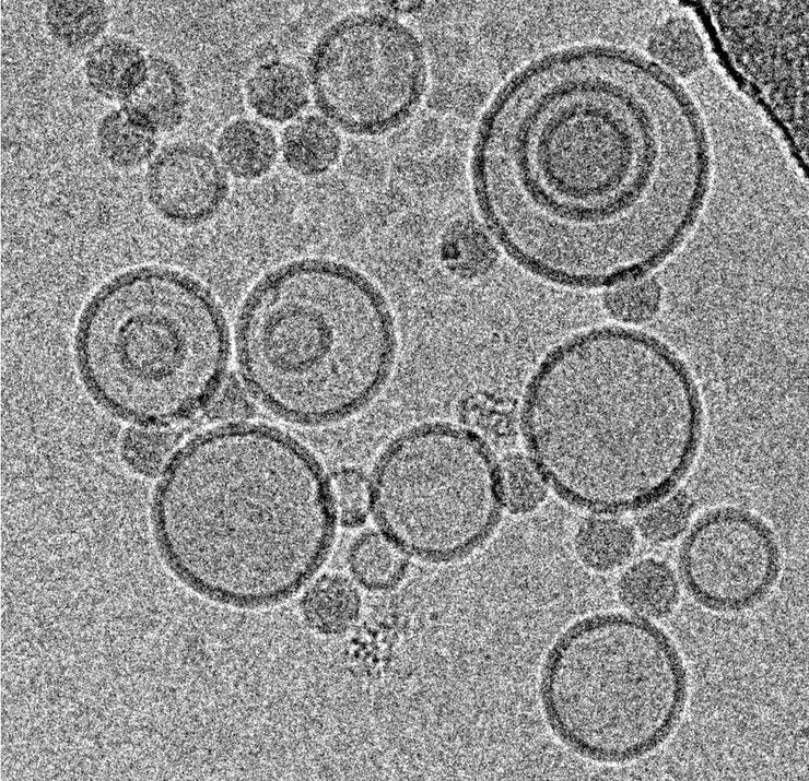
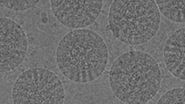
ウイルスの超微細構造の評価には、1 nm近い分解能が必要です。 現在、この分解能は電子顕微鏡によってのみ達成できます。 クライオ電子顕微鏡は、ナノメートル以下の分解能さえ達成できます。

超微細構造分解能は、宿主とウイルス間相互作用のナノスケールでの観察、異なる分析技術による結果の確認、薬物標的部位の特定を可能にします。 さらに、ウイルス粒子の相互作用時に標的となった細胞表面レセプターのリガンドや小分子は、ワクチンとウイルス侵入阻害剤の構造情報に基づいたデザインにとって鍵となります。

従来の電子顕微鏡やクライオ電子顕微鏡にとって、試料作製は極めて重要です。 ライカマイクロシステムズは、専門的な機器や知識が求められるこのプロセスを、電子顕微鏡試料作製の広範なポートフォリオでサポートします。さらに、ライカのエキスパートがアドバイスを提供します。

Related articles

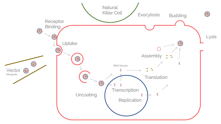
Exploring the Structure and Life Cycle of Viruses

The SARS-CoV-2 outbreak started in late December 2019 and has since reached a global pandemic, leading to a worldwide battle against COVID-19. The ever-evolving electron microscopy methods offer a…

Immersion Freezing for Cryo-Transmission Electron Microscopy: Applications

A well established usage case for cryo-TEM is three-dimensional reconstruction of isolated macromolecules, virus particles, or filaments. On one hand, these approaches are based on averaging of…
Scroll to top