
Filter articles
标签
Story Type
Products
Loading...

Applying AI and Machine Learning in Microscopy and Image Analysis
Prof. Emma Lundberg is a professor in cell biology proteomics at KTH Royal Institute of Technology, Sweden. She is also the director of the Cell Atlas, an integral part of the Swedish-based Human…
Loading...
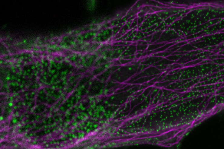
 enhanced with Aivia’s Pixel Classifier (right)")
Simplifying the Cancer Biology Image Analysis Workflow
As cancer biology data sets grow, so do the challenges in microscopy image analysis. Aivia experts cover how to overcome these challenges with AI.
Loading...

Examining Critical Developmental Events in High-Definition
Extended live cell imaging of embryo development requires a delicate balance between light exposure, temporal resolution and spatial resolution to maintain cells’ viability. Compromises between the…
Loading...

Observing Complex Cellular Interactions at Multiple Scales
Learn how to observe challenging cellular interactions with easy to deploy object detection and relationship measurements.
Loading...

Accelerating Neuron Image Analysis with Automation
The ability to examine complex neural processes relies on the accurate reconstruction of neuronal networks at scale. Most data extraction methods in neuroscience research are time-consuming and…
Loading...
Tracking Single Cells Using Deep Learning
AI-based solutions continue to gain ground in the field of microscopy. From automated object classification to virtual staining, machine and deep learning technologies are powering scientific…
Loading...

Learning the Cellular Architecture from its Optical Properties
In the last 3 years, microscopists have started to use "AI based" solutions for a wide range of applications, including image acquisition optimization (smart microscopy), object classification, image…
Loading...
AI in Microscopy Webinar
We demonstrate residual channel attention networks for restoring and enhancing volumetric time-lapse (4D) fluorescence microscopy data.
