
Filter articles
标签
Story Type
Products
Loading...

How to Prepare your Specimen for Immunofluorescence Microscopy
Immunofluorescence (IF) is a powerful method for visualizing intracellular processes, conditions and structures. IF preparations can be analyzed by various microscopy techniques (e.g. CLSM,…
Loading...
 in 3D")
How Artificial Intelligence Enhances Confocal Imaging
In this article, we show how artificial intelligence (AI) can enhance your imaging experiments. Namely, how Dynamic Signal Enhancement powered by Aivia improves image quality while capturing the…
Loading...

Spectroscopic Evaluation of Red Blood Cells
Hemoglobinopathies are a major healthcare problem. This study presents a possible diagnostic tool for thalassemia which is based on confocal spectroscopy. This approach exploits spectral detection and…
Loading...
Visualizing Protein Degradation and Aggregation in the Living Cell
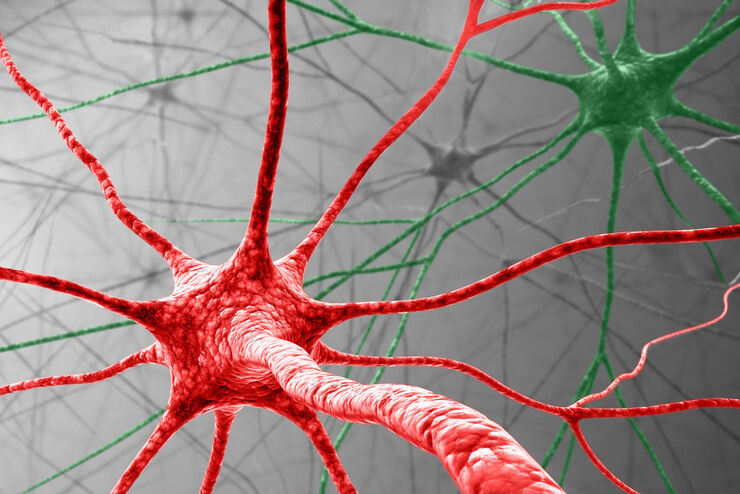
Our guest speaker, Prof Dr Eric Reits, presents his work on neurodegenerative disorders. Reits’ group are experts on the subject of Huntington’s disease and work towards identifying leads for…
Loading...
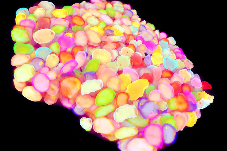
Life Beyond the Pixels: Deep Learning Methods for Single Cell Analysis
Our guest speaker Prof Dr Peter Horvath presents his work on single cell-based large-scale microscopy experiments. This novel targeting approach includes the use of machine learning models and…
Loading...

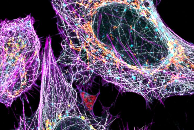
Live Cell Imaging Gallery
Live cell microscopy techniques are fundamental to get a better understanding of cellular and molecular function. Today, widefield microscopy is the most common technique used to visualize cell…
Loading...

Tissue Image Gallery
Visual analysis of animal and human tissues is critical to understand complex diseases such as cancer or neurodegeneration. From basic immunohistochemistry to intravital imaging, confocal microscopy…
Loading...
Multicolor Image Gallery
Fluorescence multicolor microscopy, which is one aspect of multiplex imaging, allows for the observation and analysis of multiple elements within the same sample – each tagged with a different…
Loading...

Cancer Research Image Gallery
Fluorescence microscopy allows the study of changes occurring in tissue and cells during cancer development and progression. Techniques such as live cell imaging are critical to understand cancer…
