
Filter articles
标签
Story Type
Products
Loading...

Fast, High-quality Vitrification with the EM ICE High Pressure Freezer
The EM ICE High Pressure Freezer was developed with a unique freezing principle and uses only a single pressurization and cooling liquid: liquified nitrogen (LN2). This design enables three major…
Loading...

Targeting Active Recycling Nuclear Pore Complexes using Cryo Confocal Microscopy
In this article, how cryo light microscopy and, in particular cryo confocal microscopy, is used to improve the reliability of cryo EM workflows is described. The quality of the EM grids and samples is…
Loading...
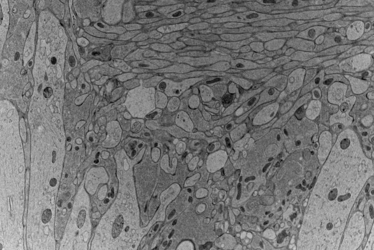
Investigating Synapses in Brain Slices with Enhanced Functional Electron Microscopy
A fundamental question of neuroscience is: what is the relationship between structural and functional properties of synapses? Over the last few decades, electrophysiology has shed light on synaptic…
Loading...
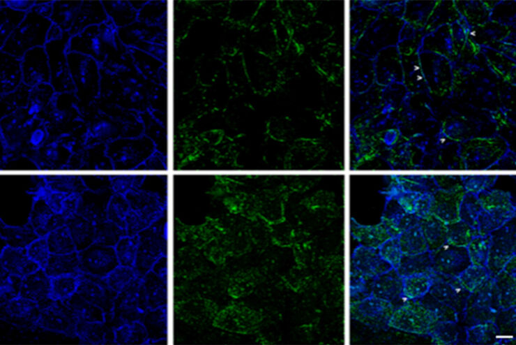
Improvement of Imaging Techniques to Understand Organelle Membrane Cell Dynamics
Understanding cell functions in normal and tumorous tissue is a key factor in advancing research of potential treatment strategies and understanding why some treatments might fail. Single-cell…
Loading...

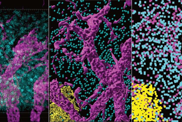
Advancing Cell Biology with Cryo-Correlative Microscopy
Correlative light and electron microscopy (CLEM) advances biological discoveries by merging different microscopes and imaging modalities to study systems in 4D. Combining fluorescence microscopy with…
Loading...

Consumables for Laser Microdissection
There are many different types of consumables for laser microdissection (LMD) systems. They cover a wide range of applications from basic to highly specialized, enabling scientists to choose their own…
Loading...

How FLIM Microscopy Helps to Detect Microplastic Pollution
The use of autofluorescence in biological samples is a widely used method to gain detailed knowledge about systems or organisms. This property is not only found in biological systems, but also…
Loading...
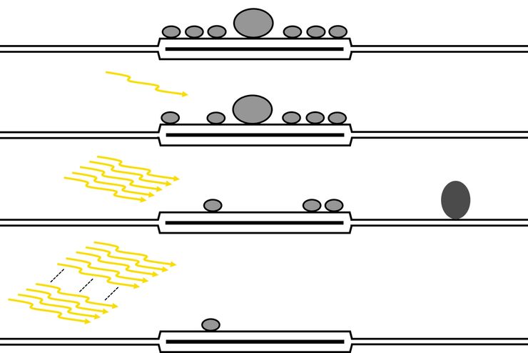
High-pressure freezing: Revealing functional mechanisms of synaptic transmission
Learn more about applying optogenetic stimulation in the EM ICE and how this technology has the potential to reveal structural and functional mechanisms of synaptic transmission. Get a detailed…
Loading...
From Organs to Tissues to Cells: Analyzing 3D Specimens with Widefield Microscopy
Obtaining high-quality data and images from thick 3D samples is challenging using traditional widefield microscopy because of the contribution of out-of-focus light. In this webinar, Falco Krüger…
