STELLARIS CRS
Microscópios confocais
Produtos
Página inicial
Leica Microsystems
STELLARIS CRS Microscópio de dispersão coerente de Raman
Explore a microscopia química livre de marcadores
Leia os nossos artigos mais recentes
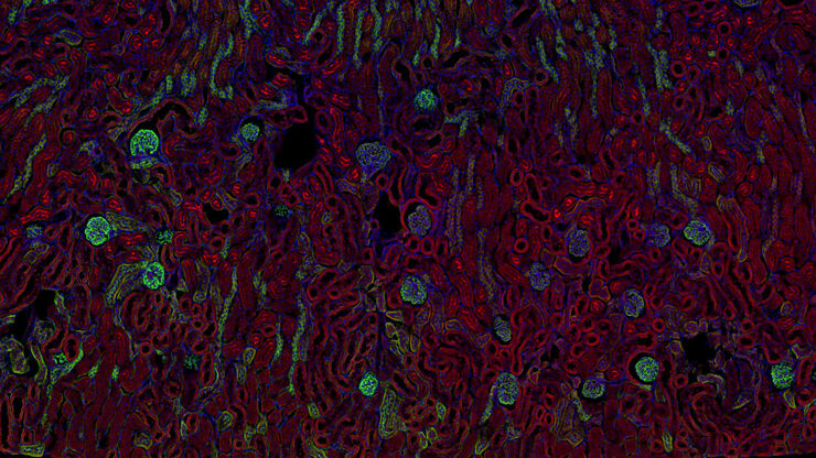
Pesquisa de câncer
O câncer é uma doença complexa e heterogênea causada por células com deficiência na regulação do crescimento. Mudanças genéticas e epigenéticas em uma ou em um grupo de células prejudicam o…
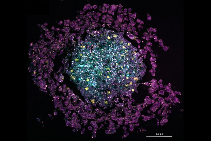
Organismos Modelo na Pesquisa
Um organismo modelo é uma espécie usada pelos pesquisadores para estudar processos biológicos específicos. Eles têm características genéticas similares aos seres humanos e são usados tipicamente em…
Neurocientífica
Você está trabalhando rumo a uma melhor compreensão de doenças neurodegenerativas ou estudando a função do sistema nervoso? Veja como você pode fazer descobertas com as soluções de aquisição de…
How to Prepare Samples for Stimulated Raman Scattering (SRS) imaging
Find here guidelines for how to prepare samples for stimulated Raman scattering (SRS), acquire images, analyze data, and develop suitable workflows. SRS spectroscopic imaging is also known as SRS…
Coherent Raman Scattering Microscopy Publication List
CRS (Coherent Raman Scattering) microscopy is an umbrella term for label-free methods that image biological structures by exploiting the characteristic, intrinsic vibrational contrast of their…
The Potential of Coherent Raman Scattering Microscopy at a Glance
Coherent Raman scattering microscopy (CRS) is a powerful approach for label-free, chemically specific imaging. It is based on the characteristic intrinsic vibrational contrast of molecules in the…
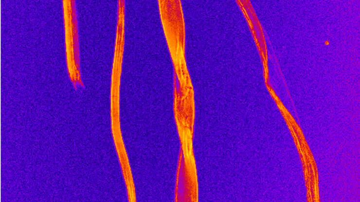
Formulated Product Characterization with SRS Microscopy
Creams, pastes, gels, emulsions, and tablets are ubiquitous across a wide range of manufacturing sectors from pharmaceuticals and consumer health products to agrochemicals and paint. To improve…
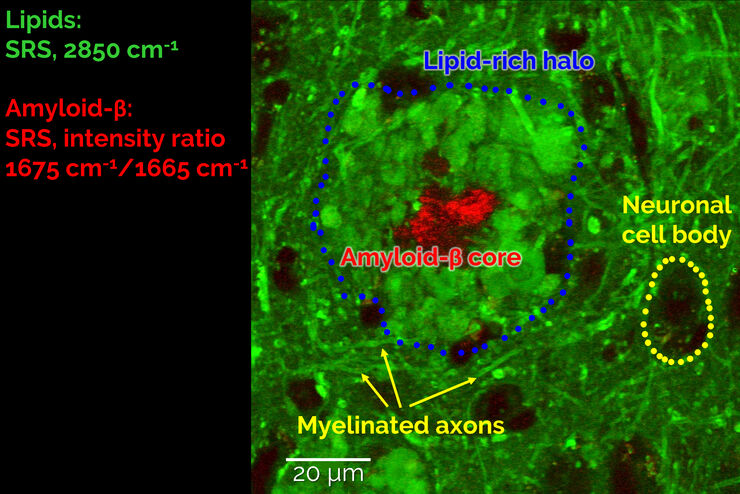
Stimulated Raman Scattering Microscopy Probes Neurodegenerative Disease
Despite decades of research, the molecular mechanisms underlying some of the most severe neurodegenerative diseases, such as Alzheimer’s or Parkinson’s, remain poorly understood. The progression of…
CARS Microscopy: Imaging Characteristic Vibrational Contrast of Molecules
Coherent anti-Stokes Raman scattering (CARS) microscopy is a technique that generates images based on the vibrational signatures of molecules. This imaging methods does not require labeling, yet…
An Introduction to CARS Microscopy
CARS overcomes the drawbacks of conventional staining methods by the intrinsic characteristics of the method. CARS does not require labeling because it is highly specific to molecular compounds which…
