
Science Lab
Science Lab
Bem-vindo ao portal de conhecimento da Leica Microsystems. Você encontrará pesquisas científicas e material didático sobre o tema microscopia. O portal oferece suporte a iniciantes, profissionais experientes e cientistas em seus trabalhos e experimentos diários. Explore tutoriais interativos e notas de aplicação, descubra os fundamentos da microscopia, bem como as tecnologias de ponta. Faça parte da comunidade do Science Lab e compartilhe sua experiência.
Filter articles
Tags
Story Type
Products
Loading...

How does Real-Time OCT Imaging Impact Precision in Corneal Surgery?
Corneal surgery is a highly specialized field. It requires great surgical precision to overcome challenges such as visualizing clearly the full anterior chamber, performing Descemet membrane peeling…
Loading...

How to Achieve Brain Tissue Resection with GLOW400 AR
Intraoperative MRI is one form of real-time intraoperative visualization, but if more in-depth visualization to identify a tumor during surgery is wanted, intraoperative fluorescence diagnostics is…
Loading...

How to Automatically Obtain Fluorescent Cells of Interest in a Block-face
Block-face created by automatic trimming under fluorescence.
Mammalian cells of interest, stained with CellTrackerTM Green are visualized within the block-face using the UC Enuity equipped with the…
Loading...

Deep Visual Proteomics Provides Precise Spatial Proteomic Information
Despite the availability of imaging methods and mass spectroscopy for spatial proteomics, a key challenge that remains is correlating images with single-cell resolution to protein-abundance…
Loading...

What is Empty Magnification and How can Users Avoid it
The phenomenon of “empty magnification”, which can occur while using an optical, light, or digital microscope, and how it can be avoided is explained in this article. The performance of an optical…
Loading...

How to Study Gene Regulatory Networks in Embryonic Development
Join Dr. Andrea Boni by attending this on-demand webinar to explore how light-sheet microscopy revolutionizes developmental biology. This advanced imaging technique allows for high-speed, volumetric…
Loading...

Spatial Analysis of Neuroimmune Interactions in Alzheimer’s Disease
Alzheimer’s disease (AD) is a complex neurodegenerative disorder characterized by neurofibrillary tangles, β-amyloid plaques, and neuroinflammation. These dysfunctions trigger or are exacerbated by…
Loading...
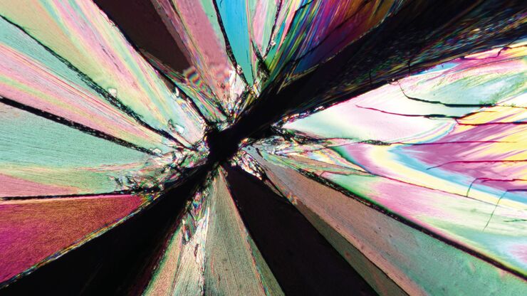
The Polarization Microscopy Principle
Polarization microscopy is routinely used in the material and earth sciences to identify materials and minerals on the basis of their characteristic refractive properties and colors. In biology,…
Loading...

A Guide to Spatial Biology
What is spatial biology, and how can researchers leverage its tools to meet the growing demands of biological questions in the post-omics era? This article provides a brief overview of spatial biology…
