
Ciências da vida
Ciências da vida
Este é o lugar para expandir seus conhecimentos, recursos de pesquisa e aplicações práticas de microscopia em vários campos científicos. Saiba como obter visualização precisa, interpretação de imagens e avanços na pesquisa. Encontre informações perspicazes sobre microscopia avançada, técnicas de geração de imagens, preparação de amostras e análise de imagens. Os tópicos abordados incluem biologia celular, neurociência e pesquisa do câncer, com foco em aplicações e inovações de ponta.
Filter articles
Tags
Story Type
Products
Loading...

RNA Quality after Different Tissue Sample Preparation
The influence of sample preparation and ultraviolet (UV) laser microdissection (UV LMD) on the quality of RNA from murine-brain tissue cryo-sections is described in this article. To obtain good…
Loading...

New Imaging Tools for Cryo-Light Microscopy
New cryo-light microscopy techniques like LIGHTNING and TauSense fluorescence lifetime-based tools reveal structures for cryo-electron microscopy.
Loading...
 in 14 x 18 tiles. Lifetime gives an additional contrast that allows to differentiate different structures in histological stainings.")
A Guide to Fluorescence Lifetime Imaging Microscopy (FLIM)
The fluorescence lifetime is a measure of how long a fluorophore remains on average in its excited state before returning to the ground state by emitting a fluorescence photon.
Loading...
, the cis-golgi matrix protein GM130 (AF488, green), and the trans-golgi network membrane protein TGN46 (AF647, red).")
Golgi Organizational Changes in Response to Cell Stress
In this video on demand, our special guest George Galea from EMBL Heidelberg will look at HeLa Kyoto cells treated with various chemotherapeutic agents to investigate their effect on the Golgi complex…
Loading...

Multiplexed Imaging Types, Benefits and Applications
Multiplexed imaging is an emerging and exciting way to extract information from human tissue samples by visualizing many more biomarkers than traditional microscopy. By observing many biomarkers…
Loading...

3D Tissue Imaging: From Fast Overview To High Resolution With One Click
3D Tissue imaging is a widespread discipline in the life sciences. Researchers use it to reveal detailed information of tissue composition and integrity, to make conclusions from experimental…
Loading...

How To Perform Fast & Stable Multicolor Live-Cell Imaging
With the help of live-cell imaging researchers gain insights into dynamic processes of living cells up to whole organisms. This includes intracellular as well as intercellular activities. Protein or…
Loading...

Imaging of Cardiac Tissue Regeneration in Zebrafish
Learn how to image cardiac tissue regeneration in zebrafish focusing on cell proliferation and response during recovery with Laura Peces-Barba Castaño from the Max Planck Institute.
Loading...
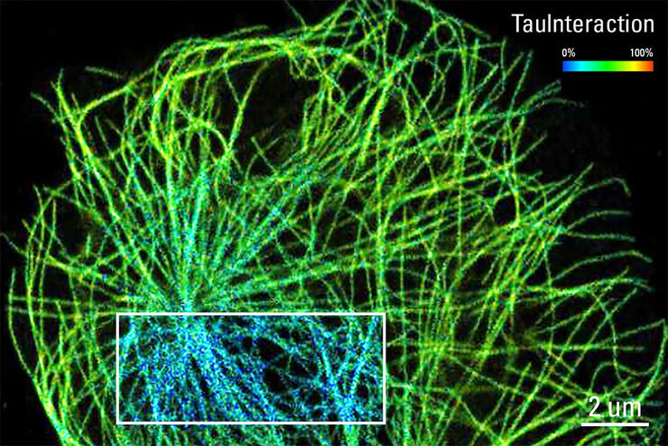
TauInteraction – Studying Molecular Interactions with TauSense
Fluorescence microscopy constitutes one of the pillars in life sciences and is a tool commonly used to unveil cellular structure and function. A key advantage of fluorescence microscopy resides in the…
