
is mobile? false
Ciências da vida
Ciências da vida
Este é o lugar para expandir seus conhecimentos, recursos de pesquisa e aplicações práticas de microscopia em vários campos científicos. Saiba como obter visualização precisa, interpretação de imagens e avanços na pesquisa. Encontre informações perspicazes sobre microscopia avançada, técnicas de geração de imagens, preparação de amostras e análise de imagens. Os tópicos abordados incluem biologia celular, neurociência e pesquisa do câncer, com foco em aplicações e inovações de ponta.
Popular Show subnavigation
Ciências da vida


Filter articles
Tags
Story Type
Products
Loading...

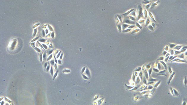
How to Improve Live Cell Imaging with Coral Life
For live-cell CLEM applications, light microscopy imaging is a critical step for identifying the right cell in the right state at the right time. In this article, Leica experts share their insights on…
Loading...

How to Keep Your Samples Under Physiological Conditions
The Coral Life workflow combines dynamic data with the best possible sample fixation by high pressure freezing. However, good sample preservation won’t help if your cells are stressed by temperature…
Loading...

Fast, High-quality Vitrification with the EM ICE High Pressure Freezer
The EM ICE High Pressure Freezer was developed with a unique freezing principle and uses only a single pressurization and cooling liquid: liquified nitrogen (LN2). This design enables three major…
Loading...

Targeting Active Recycling Nuclear Pore Complexes using Cryo Confocal Microscopy
In this article, how cryo light microscopy and, in particular cryo confocal microscopy, is used to improve the reliability of cryo EM workflows is described. The quality of the EM grids and samples is…
Loading...
 from the desired fluorescent signal from the cell membrane (green).")
Learn how to Remove Autofluorescence from your Confocal Images
Autofluorescence can significantly reduce what you can see in a confocal experiment. This article explores causes of autofluorescence as well as different ways to remove it, from simple media fixes to…
Loading...

Bridging Structure and Dynamics at the Nanoscale through Optogenetics and Electrical Stimulation
Nanoscale ultrastructural information is typically obtained by means of static imaging of a fixed and processed specimen. However, this is only a snapshot of one moment within a dynamic system in…
