
Science Lab
Science Lab
Bem-vindo ao portal de conhecimento da Leica Microsystems. Você encontrará pesquisas científicas e material didático sobre o tema microscopia. O portal oferece suporte a iniciantes, profissionais experientes e cientistas em seus trabalhos e experimentos diários. Explore tutoriais interativos e notas de aplicação, descubra os fundamentos da microscopia, bem como as tecnologias de ponta. Faça parte da comunidade do Science Lab e compartilhe sua experiência.
Filter articles
Tags
Story Type
Products
Loading...

An Introduction to Computational Clearing
Many software packages include background subtraction algorithms to enhance the contrast of features in the image by reducing background noise. The most common methods used to remove background noise…
Loading...
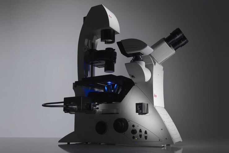
Factors to Consider When Selecting a Research Microscope
An optical microscope is often one of the central devices in a life-science research lab. It can be used for various applications which shed light on many scientific questions. Thereby the…
Loading...

Challenges Faced When Manually Rating Non-Metallic Inclusions (NMIs) to Determine Steel Quality
Rapid, accurate, and reliable rating of non-metallic inclusions (NMIs) is instrumental for the determination of steel quality. This article describes the challenges that arise from manual NMI rating,…
Loading...
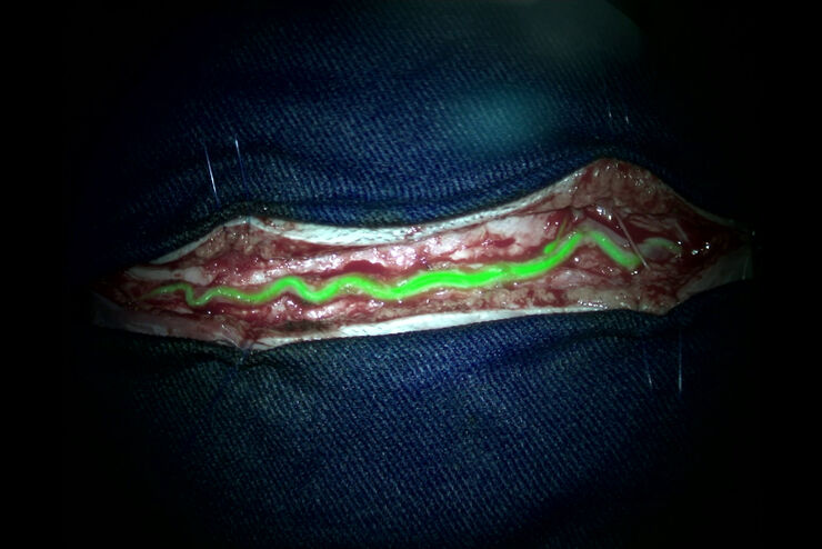
 - THUNDER Imager 3D Cell Culture Influenca virus – red, cilia – green, Nuclei – blue.")
How Can Immunofluorescence Aid Virology Research?
Modern virology research has become as crucial now as ever before due to the global COVID-19 pandemic. There are many powerful technologies and assays that virologists can apply to their research into…
Loading...
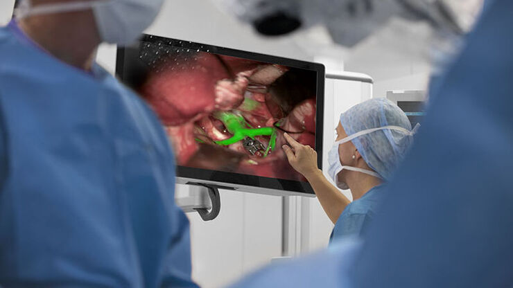
Augmented Reality (AR) Fluorescence Image Gallery
Building on a decade of leadership in fluorescence imaging technology, GLOW800 AR fluorescence is the first of many modalities based on the proprietary GLOW AR platform.
The sophisticated imaging…
Loading...
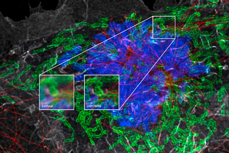
Obtenha o máximo de informações de sua amostra com o LIGHTNING
O LIGHTNING é um processo adaptivo totalmente automático para a extração de informações que revela estruturas finas e detalhes que, de outra forma, simplesmente não seriam visíveis. Ao contrário de…
Loading...
Advanced Techniques in Cataract and Refractive Surgery
In this webinar Dr. Thompson and Dr. Moshirfar will explain how Leica microscopes aid in procedures such as Centration of Multifocal IOLs and corneal inlays such as Kamra and Lenticular Grafts used in…
Loading...
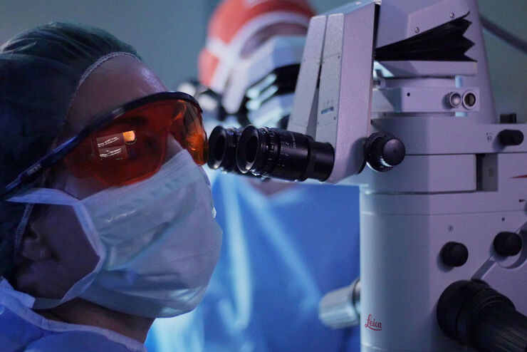
Clinical Uses in Cerebrovascular and Skull Base Neurosurgery
In this webinar Dr. Bendok and Dr. Morcos explain how Augmented Reality and Fluorescence can enhance visualization and support surgical decision making. They present first-hand experience of the GLOW…
Loading...
What is intraoperative OCT telling us?
In the following videos are excerpts taken from a webinar delivered by Dr. Barbara Parolini. During the webinar Dr. Parolini discusses and illustrates how intraoperative OCT provides real-time,…
