
Science Lab
Science Lab
Bem-vindo ao portal de conhecimento da Leica Microsystems. Você encontrará pesquisas científicas e material didático sobre o tema microscopia. O portal oferece suporte a iniciantes, profissionais experientes e cientistas em seus trabalhos e experimentos diários. Explore tutoriais interativos e notas de aplicação, descubra os fundamentos da microscopia, bem como as tecnologias de ponta. Faça parte da comunidade do Science Lab e compartilhe sua experiência.
Filter articles
Tags
Story Type
Products
Loading...

Cataract Surgery with CoAx4 Illumination
A stable red reflex is one of the most important features of an ophthalmic surgical microscope for cataract surgery. It’s the red reflex that makes the structure of the lens visible and thus makes for…
Loading...
")
A Brief History of Light Microscopy
The history of microscopy begins in the Middle Ages. As far back as the 11th century, plano-convex lenses made of polished beryl were used in the Arab world as reading stones to magnify manuscripts.…
Loading...

From Light to Mind: Sensors and Measuring Techniques in Confocal Microscopy
This article outlines the most important sensors used in confocal microscopy. By confocal microscopy, we mean "True Confocal Scanning", i.e. the technique that illuminates and measures one single…
Loading...
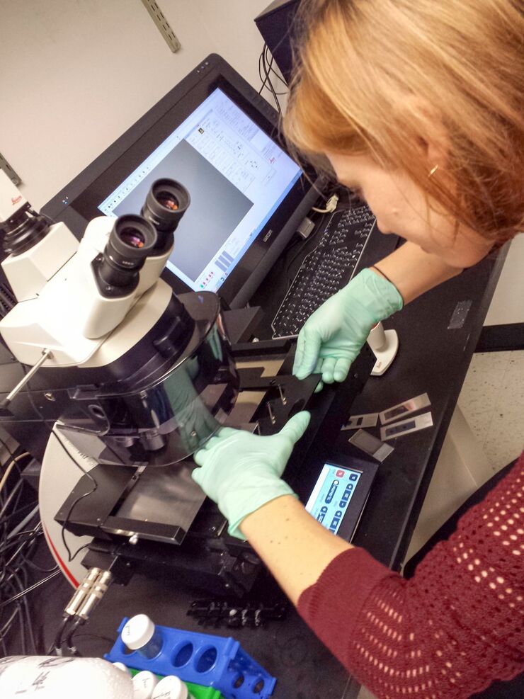
Workflows & Protocols: How to Use a Leica Laser Microdissection System and Qiagen Kits for Successful RNA Analysis
Laser Microdissection (LMD) allows isolating individual cells or chromosomes and is a well established technique for sample preparation prior downstream analysis of the nucleic acid content via PCR or…
Loading...

Confocal and Light Sheet Imaging
Optical imaging instrumentation can magnify tiny objects, zoom in on distant stars and reveal details that are invisible to the naked eye. But it notoriously suffers from an annoying problem: the…
Loading...

Universal PAINT – Dynamic Super-Resolution Microscopy
Super-resolution microscopy techniques have revolutionized biology for the last ten years. With their help cellular components can now be visualized at the size of a protein. Nevertheless, imaging…
Loading...
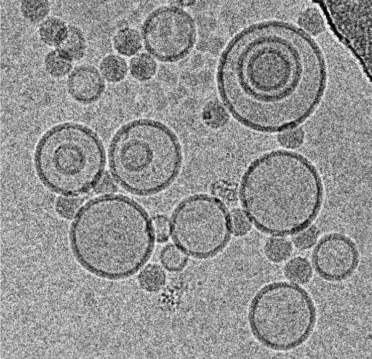
Immersion Freezing for Cryo-Transmission Electron Microscopy: Applications
A well established usage case for cryo-TEM is three-dimensional reconstruction of isolated macromolecules, virus particles, or filaments. On one hand, these approaches are based on averaging of…
Loading...
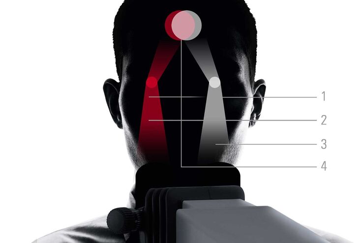
FusionOptics in Neurosurgery and Ophthalmology – for a Larger 3D Area in Focus
Neurosurgeons and ophthalmologists deal with delicate structures, deep or narow cavities and tiny structures with vitally important functions. A clear, three-dimensional view on the surgical field is…
Loading...
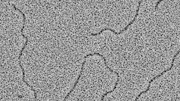
Glycerol Spraying/Platinum Low Angle Rotary Shadowing of DNA with the Leica EM ACE600 e-beam
Glycerol spraying/low angle rotary shadowing (Aebi and Baschong, 2006) is a preparation technique used in biology to visualize structures yielding insufficent contrast with other techniques, due to…
